A. AP KUB (Fig. 3.4) Description of possible error: 1. Anatomy demonstrated: 2. Part positioning: 3. Collimation and central ray: 4. Exposure: 5. Anatomic side markers: Repeatable error(s): 3. AP erect abdomen (Fig. 3.5) Description of possible error: 1. Anatomy demonstrated: 2. Part positioning: 3. Collimation and central ray: 4. Exposure: 5. Anatomic side markers: Repeatable error(s): DE 10 Fig. 3.4 Anteroposterior image of kidneys, ureters, and bladder. (Case courtesy of Dr. Jeremy Jones, Radiopaedia.org, rID: 34067.) Erect Ⓒ Fig. 3.5 Anteroposterior image of erect abdomen.
A. AP KUB (Fig. 3.4) Description of possible error: 1. Anatomy demonstrated: 2. Part positioning: 3. Collimation and central ray: 4. Exposure: 5. Anatomic side markers: Repeatable error(s): 3. AP erect abdomen (Fig. 3.5) Description of possible error: 1. Anatomy demonstrated: 2. Part positioning: 3. Collimation and central ray: 4. Exposure: 5. Anatomic side markers: Repeatable error(s): DE 10 Fig. 3.4 Anteroposterior image of kidneys, ureters, and bladder. (Case courtesy of Dr. Jeremy Jones, Radiopaedia.org, rID: 34067.) Erect Ⓒ Fig. 3.5 Anteroposterior image of erect abdomen.

Related questions
Question
Transcribed Image Text:A. AP KUB (Fig. 3.4)
Description of possible error:
1. Anatomy demonstrated:
2. part positioning:
3. Collimation and central ray:
4. Exposure:
5.
Anatomic side markers:
Repeatable error(s):
B. AP erect abdomen (Fig. 3.5)
Description of possible error:
1. Anatomy demonstrated:
2. Part positioning:
3. Collimation and central ray:
4. Exposure:
5. Anatomic side markers:
Repeatable error(s):
liw
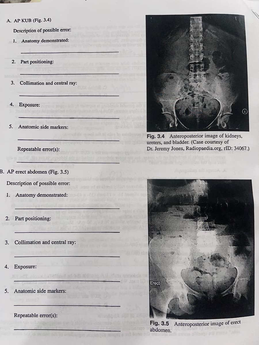
Fig. 3.4 Anteroposterior image of kidneys,
ureters, and bladder. (Case courtesy of
Dr. Jeremy Jones, Radiopaedia.org, rID: 34067.)
DE 10 Erect
Fig. 3.5 Anteroposterior image of erect
nos abdomen.
Expert Solution
This question has been solved!
Explore an expertly crafted, step-by-step solution for a thorough understanding of key concepts.

This is a popular solution!

Trending now
This is a popular solution!

Step by step
Solved in 3 steps
