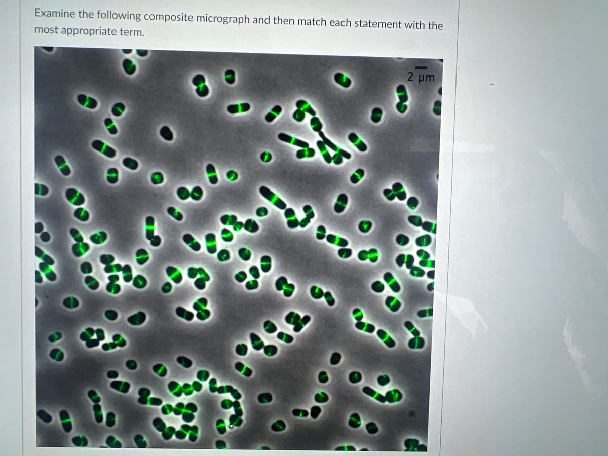
e fluorescent label in this image is he cell shapes are imaged with The microscopy used for determining he localization of proteins is he cellular process labelled in green is Electron microscopy The protein visualized in this image is The cell morphology is The appoximate size of the cells is three [Choose ] [Choose ] DNA replication ◄ Previous Nanometers Phase-contrast microscopy Actin Heterotrophic Pleomorphic Cell morphogenesis Microns Binary fission Primary antibodies FtsZ Polymorphic Globular fusion protein Fluorescence microscopy DNA-GFP Green fluorescent protein No Dark-field microscopy


DNA and RNA
Deoxyribonucleic acid (DNA) is usually called the blueprint of life. Deoxyribose is a monosaccharide that has a key function in the synthesis of deoxyribonucleic acid. One less oxygen-containing hydroxyl group occurs in deoxyribose sugar. Nucleic acid, deoxyribonucleic acid, is one of the natural components. Deoxyribonucleic acid is a double-stranded molecule. Watson and Crick postulated the double-stranded model of the helix. A deoxyribonucleic acid is a molecular group that carries and transmits genetic information from parents to offspring. All eukaryotic and prokaryotic cells are involved.
DNA as the Genetic Material
DNA, or deoxyribonucleic acid, is a long polymeric nucleic acid molecule discovered in the late 1930s. It is a polymer; a long chain-like molecule made up of several monomers connected in a sequence. It possesses certain characteristics that qualify it as a genetic component. Certain organisms have different types of nucleic acids as their genetic material - DNA or RNA.
Genetics
The significant branch in science which involves the study of genes, gene variations, and the organism's heredity is known as genetics. It is also used to study the involvement of a gene or set of genes in the health of an individual and how it prevents several diseases in a human being. Thus, genetics also creates an understanding of various medical conditions.
DNA Replication
The mechanism by which deoxyribonucleic acid (DNA) is capable of producing an exact copy of its own is defined as DNA replication. The DNA molecules utilize a semiconservative method for replication.
![The fluorescent label in this image is
The cell shapes are imaged with
The microscopy used for determining
the localization of proteins is
The cellular process labelled in green is Electron microscopy
The protein visualized in this image is
The cell morphology is
The appoximate size of the cells is
three
[Choose ]
[Choose ]
DNA replication
◄ Previous
Nanometers
Phase-contrast microscopy
Actin
Heterotrophic
Pleomorphic
Cell morphogenesis
Microns
Binary fission
Primary antibodies
FtsZ
Polymorphic
Globular fusion protein
Fluorescence microscopy
DNA-GFP
Green fluorescent protein
No Dark-field microscopy
Peptone
d at 12](/v2/_next/image?url=https%3A%2F%2Fcontent.bartleby.com%2Fqna-images%2Fquestion%2F14003164-91a4-4641-b129-b94e9b7a2acb%2F4ace6509-8b38-41b9-8d03-b4e125d49fba%2Fhhkal0a_processed.jpeg&w=3840&q=75)

Step by step
Solved in 3 steps


