
Surgical Tech For Surgical Tech Pos Care
5th Edition
ISBN:9781337648868
Author:Association
Publisher:Association
Chapter2: Legal Concepts, Risk Management, And Ethical Issues
Section: Chapter Questions
Problem 2.4CS

Related questions

Question
read the article that is in the images and write the most important data
Transcribed Image Text:11:51 p. m. Mar 28 mar.
190
web.whatsapp.com
PART TWO Radiographic Positioning and Related Anatomy
Further Views
In case of injury, for gastric contrast studies, or with concern about fluid or free air, other views can be taken.
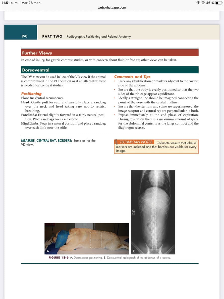
Dorsoventral
The DV view can be used in lieu of the VD view if the animal
is compromised in the VD position or if an alternative view
is needed for contrast studies.
Positioning
Place In: Ventral recumbency.
Head: Gently pull forward and carefully place a sandbag
over the neck and head taking care not to restrict
breathing.
Forelimbs: Extend slightly forward in a fairly natural posi-
tion. Place sandbags over each elbow.
Hind Limbs: Keep in a natural position, and place a sandbag
over each limb near the stifle.
MEASURE, CENTRAL RAY, BORDERS: Same as for the
VD view.
Comments and Tips
• Place any identification or markers adjacent to the correct
side of the abdomen.
• Ensure that the body is evenly positioned so that the two
sides of the rib cage appear equidistant.
•
Ideally a straight line should be imagined connecting the
point of the nose with the caudal midline.
• Ensure that the sternum and spine are superimposed; the
image receptor and central ray are perpendicular to both.
• Expose immediately at the end phase of expiration.
During expiration there is a maximum amount of space
for the abdominal contents as the lungs contract and the
diaphragm relaxes.
TECHNICIAN NOTES Collimate; ensure that labels/
markers are included and that borders are visible for every
image.
Я
B
FIGURE 18-6 A, Dorsoventral positioning. B, Dorsoventral radiograph of the abdomen of a canine.
46%
Transcribed Image Text:11:51 p. m. Mar 28 mar.
252 de 633
web.whatsapp.com
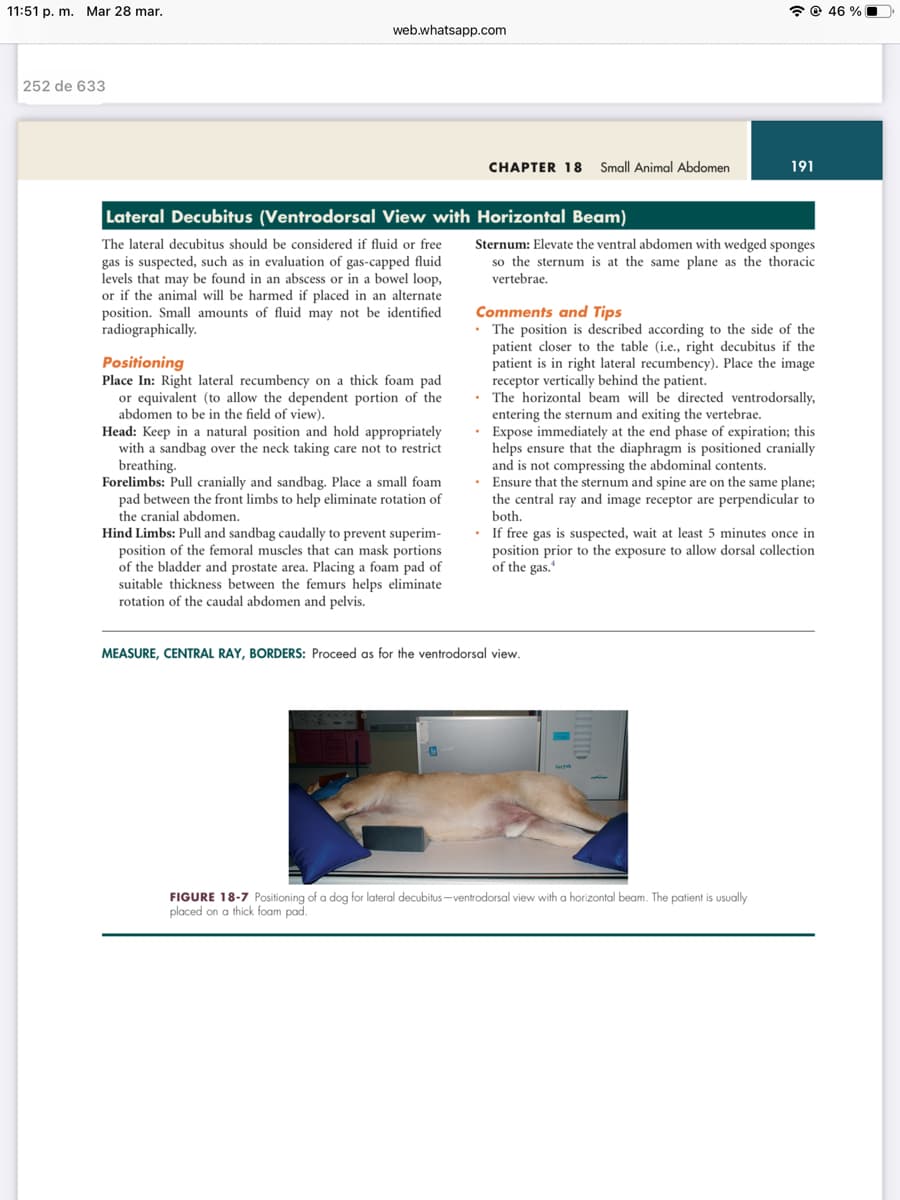
Lateral Decubitus (Ventrodorsal View with
The lateral decubitus should be considered if fluid or free
gas is suspected, such as in evaluation of gas-capped fluid
levels that may be found in an abscess or in a bowel loop,
or if the animal will be harmed if placed in an alternate
position. Small amounts of fluid may not be identified
radiographically.
Positioning
Place In: Right lateral recumbency on a thick foam pad
or equivalent (to allow the dependent portion of the
abdomen to be in the field of view).
Head: Keep in a natural position and hold appropriately
with a sandbag over the neck taking care not to restrict
breathing.
Forelimbs: Pull cranially and sandbag. Place a small foam
pad between the front limbs to help eliminate rotation of
the cranial abdomen.
Hind Limbs: Pull and sandbag caudally to prevent superim-
position of the femoral muscles that can mask portions
of the bladder and prostate area. Placing a foam pad of
suitable thickness between the femurs helps eliminate
rotation of the caudal abdomen and pelvis.
CHAPTER 18 Small Animal Abdomen
@46 %
Horizontal Beam)
Sternum: Elevate the ventral abdomen with wedged sponges
so the sternum is at the same plane as the thoracic
vertebrae.
191
Comments and Tips
. The position is described according to the side of the
patient closer to the table (i.e., right decubitus if the
patient is in right lateral recumbency). Place the image
receptor vertically behind the patient.
The horizontal beam will be directed ventrodorsally,
entering the sternum and exiting the vertebrae.
Expose immediately at the end phase of expiration; this
helps ensure that the diaphragm is positioned cranially
and is not compressing the abdominal contents.
• Ensure that the sternum and spine are on the same plane;
the central ray and image receptor are perpendicular to
both.
MEASURE, CENTRAL RAY, BORDERS: Proceed as for the ventrodorsal view.
If free gas is suspected, wait at least 5 minutes once in
position prior to the exposure to allow dorsal collection
of the gas.
FIGURE 18-7 Positioning of a dog for lateral decubitus-ventrodorsal view with a horizontal beam. The patient is usually
placed on a thick foam pad.
Expert Solution
This question has been solved!
Explore an expertly crafted, step-by-step solution for a thorough understanding of key concepts.

Step by step
Solved in 3 steps

Knowledge Booster


Learn more about
Need a deep-dive on the concept behind this application? Look no further. Learn more about this topic, biology and related others by exploring similar questions and additional content below.Recommended textbooks for you
Surgical Tech For Surgical Tech Pos Care
Health & Nutrition
ISBN:
9781337648868
Author:
Association
Publisher:
Cengage
Basic Clinical Lab Competencies for Respiratory C…
Nursing
ISBN:
9781285244662
Author:
White
Publisher:
Cengage
Essentials of Pharmacology for Health Professions
Nursing
ISBN:
9781305441620
Author:
WOODROW
Publisher:
Cengage

Medical Terminology for Health Professions, Spira…
Health & Nutrition
ISBN:
9781305634350
Author:
Ann Ehrlich, Carol L. Schroeder, Laura Ehrlich, Katrina A. Schroeder
Publisher:
Cengage Learning
Understanding Health Insurance: A Guide to Billin…
Health & Nutrition
ISBN:
9781337679480
Author:
GREEN
Publisher:
Cengage

Comprehensive Medical Assisting: Administrative a…
Nursing
ISBN:
9781305964792
Author:
Wilburta Q. Lindh, Carol D. Tamparo, Barbara M. Dahl, Julie Morris, Cindy Correa
Publisher:
Cengage Learning