Q: what do you think is the importance of examining the base before and after using the microscope?
A: Introduction A typical optical microscope has the following parts: Eye piece Objective lens Stage…
Q: Which part of the microscope holds the slide or specimen? the body tube the condenser the fine…
A: A microscope is an instrument used to see objects that are too small to be seen by the naked eye.…
Q: What is importance of labeling preserved specimen?
A: Labeling specimens: It is the process of naming an specimen so that it is easier to identified the…
Q: As with standard phase contrast microscopy,differential-interference contrast microscopy depends on…
A: Differential interference microscopy needs many optical parts, thus it may be terribly costly to…
Q: You are looking at a specimen on low power and you cannot see details that your instructor asked you…
A: If I am looking at a specimen at low power and can't see details that my instructor asked me to…
Q: Using a Light Microscopé to PRE-LAB QUESTIONS Fill in the diagram of the microscope with the term or…
A: A microscope is an instrument used to view objects that are not visible to the naked eye. There are…
Q: The microscopic structure pointed at is . ... the condenser lens the light source • the ocular lens…
A: Microscope is an optical instrument which has major application in visualizing very small to…
Q: A. Purpose: Figure 1 B. Materials: Microscope Magazine Slides and cover slips Paper towels Pipette…
A: purpose : to determine the total magnification power of a microscope with objectives of different…
Q: Which of these is NOT a best practice when using, handling, or storing a microscope and microscope…
A: Microscope and microscope slides should be properly handled and taken care of. In order to protect…
Q: If your object appears too small, you can increase its size on a compound microscope by altering the…
A: A microscope is the instrument by which we can observe very little object. Microscopes are of…
Q: What do you mean by deconvolution microscopy technique?
A: The organisms or substances that cannot be observed with unaided eyes are microscopic. The equipment…
Q: Why is depth of field important in studying biological structures? How can it affect your ability to…
A: Microscopy is a technique that is used to visualize objects that are not visible to the naked eyes.…
Q: She places the microscope next to a lamp. What part of the microscope can she move make sure the…
A: A microscope is a tool for examining small objects, including cells. At least one lens in the…
Q: Why should you always begin to use a microscope with a low-power objective?
A: Microscope is an instrument used to see the objects that cannot be seen by naked eye. A light…
Q: What alternative measures can be done if preservation of specimens is not possible?
A: The specimens collected has specific timeframes for these be fresh in nature. Only a fresh specimen…
Q: How do you identify which objective lens are you using to view your sample?
A: microscope objectives feature a range of magnifications from 1.25x to 150x. This is the first…
Q: you had to dispore tissue and towels from dissection? you had to dicard dispoable microscope slide…
A: The dissection is the process of dismembering the body of a deceased animal or plant for studying…
Q: Ana was tasked to measure the actual magnification of her specimen mounted on a slide without…
A: A light microscope or optical microscope is used to study a thin section of a rock. The section is…
Q: What are the ways you can control the amount of light passing through your specimen? One way is…
A: A specimen is a sample which represents the whole group. The specimen can anything which is a…
Q: A microscope is turned on, under low power, with a slide positioned on the stage ready to focus.…
A: Microscope allows the finer details of the sample placed on the slide to be viewed. As the slide in…
Q: Drag the appropriate label to the appropriate target. Reset Help Further magnifies the image and…
A:
Q: Identify and label the parts of the specimen and give a detailed description
A: The stomach is a muscular organ situated on the upper abdomen's left side. The oesophagus delivers…
Q: Which of the following microscopes allows study of living specimens? The scanning electron…
A: Microscopy is the branch of biological sciences in which the minute structures like the cells and…
Q: Does the specimen on the slide change size as you change objectives and get closer or farther out?…
A: A coumpond microscope is used to magnifying the image of any object or specimen.
Q: ewing Microscopic Objects" section to identify one way you can manipulate your slide to increase…
A: Answer: viewing microscopic objects section to identify one way you can manipulate your slide to…
Q: If you observe a specimen under this 20X objective and the specimen spans one-quarter of the field…
A: A light microscope is common equipment in most laboratories. It comes with one or two eyepieces…
Q: Why are stereomicroscopes often referred to as dissecting microscopes? Stereomicroscopes come…
A: The stereoscopic is an optical microscope variant designed for low magnification observation of a…
Q: If you are using a microscope and a specimen takes up 1/4 of the field of vview at 1000X, what is…
A: Magnification = 1000X Specimen takes 1/4 of the field of view.
Q: In case you forgot to identify and label the specimen, this can be easily troubleshooted since body…
A:
Q: Plssssss helppppp, Describe the differences you see between the three slides.
A: Thank you for the question Answer :- Abnormal blood smear 1 Shows presence of Sickle shaped cells…
Q: The specimen you are examining is very thin and transparent. What can you do to make it easier to…
A: Adjustments that we can make to the microscope to make it easier to see are:-
Q: The specimen that you are trying to find is very small and you cannot see it with scanning power.…
A: Microscopy is a field which involves using a microscope to view those specimens in enlarged size…
Q: Fill in the charts below. For your actual microscope, you will find the magnifications of the ocular…
A: Microscopy is a technique of incorporating microscopes to enlarge the size of objects (invisible to…
Q: In light microscopy, when magnification is increased, the lens focuses closer to the sample. This…
A: Microscope helps to observe the organisms or objects that cannot be seen by our naked eye because…
Q: What is the function of the microscope diaphragm? source of light condenses light from the light…
A: Microscope is an optical device that is used for observing the different objects that are invisible…
Q: Microscope type Description Magnification range (total) Image orientation Types of specimens that…
A: An instrument that is used for investigating small objects and structures is called the microscope.…
Q: Microscopic samples can be stained. the stains are used to increase and improve O resolution and…
A: Staining It is defined as a technique through which the cells or thin sections of biological tissue…
Q: is a curved glass that bends light that passes through it. regulates the amount of light on the…
A: Since we have asked to answer only one question, I am answering the first three questions of fill in…
Q: what would you do if you had to dispose tissues and towels from dissection?
A: Tissues and towels from dissection need to be safely handled to prevent infection from body fluids…
Q: Which of the following parts was absent from Leeuwenhoek’smicroscopes?a. focusing screwb. lensc.…
A: Leeuwenhoek microscope was a simple single lens device but it had greater clarity and magnification…
Q: There are two light sources in a dissecting microscope, one in the base and another in the arm.…
A: A dissecting microscope is used when you want to view the 3D structure of the specimens that are…
Q: Draw one acceptable pattern that is used in streak plate method
A: Streak plate method : It is used for the isolation into a pure culture of the organisms , f9om a…
Q: Explain why parfocal imaging is important, especially when viewing very small specimens using the…
A: Parfocal imaging involves the use of a parfocal lens that helps to keep the focus when the…
Q: Rowena was tasked to measure the actual magnification of her specimen mounted on a slide without…
A: A microscope is a piece of scientific equipment which magnifies very small objects that are too…
Q: While using a microscope, when do you use immersion oil? a. When viewing a slide under the 100x…
A: INTRODUCTION When viewing stained smears under 100x we use immersion oil. That can increase…
Q: Using a compound microscope, if I move my sample from down to up (1), in which direction will the…
A: Microscope is device used to magnify small objects and to study smaller specimen in microbiology.
Q: The girl is focusing a slide and she is moving the stage up toward the slide. a) What has happened…
A: Introduction There are various types of microscopes available in the market which have different…
The specimen you are examining is very thin and transparent. What can you do to make it easier to see
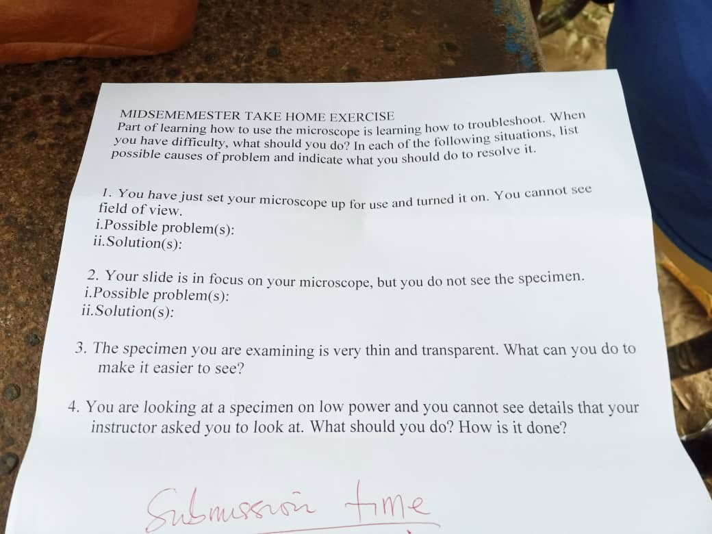
Trending now
This is a popular solution!
Step by step
Solved in 2 steps
- What information can be obtained from the slides prepared by wet mount and hanging drop technique for microscopic observation? Give at least three (3) information. What are the limitations of each method. (Provide 2 limitations)a. How was the specimen prepared for the microscopy technique applied? (for e.g. stained with H&E stain, Gram stain, unstained) b. What is the microscopy technique and magnification used to obtain this image? c. What is the basic principle of image formation using this microscopy technique? d. What can be observed and concluded from the image of the specimen? e. Are there any potential aberrations present in this specimen image? Describe these and how they may affect interpretation of the result.A medical technologist intern is preparing a blood agar medium on a hot plate while the other intern is performing an acid-fast stain (AFS) less than 1 foot away. Which of the following hazards can be most likely and immediately occur? a. Fire hazard b. Chemical hazard c. Biological hazard d. Electrical hazard
- 1. A microscope is turned on, under low power, with a slide positioned on the stage ready to focus. The specimen is visible but not in focus at all. - write the correct procedural steps used to find and focus on the specimen under medium power.Which of the following statements are true in the context of the microscope's field of view? I. It is the observable area within a circular space as defined by the lenses II. FOV is the smallest on the lowest power objective III. FOV is the maximum area visible through the microscope eyepiece IV. When you switch to a higher power, the field of zooms out towards the centerWhich of the follow are correct statements about microscope handling and care? 1. Always use TWO HANDS to carry the microscope. 2. Only use LENS PAPER to clean the lenses. Never wipe back and forth, but simply make a gentle swipe across the lens. 3. A soft cloth should be used to clean the lenses. 4. Start and finish the day’s work at the LOWEST MAGNIFICATION. 5. Start and finish the day’s work at the HIGHEST MAGNIFICATION 6. Always BEGIN examining a specimen with a LOW POWER objective. 7. Always BEGIN examining a specimen with a HIGHEST POWER objective 8. On the compound microscope, NEVER use the coarse adjustment knob with the high power or oil objective. 9. On the compound microscope, ALWAYS use the coarse adjustment knob with the high power or oil objective. 10. Unplug the microscope by pulling on the plug, not the cord.
- The following questions are based upon viewing a sample (the letter “e”) under a microscope with lower power objective 1) Does contrast increased or decreased as the amount of light incident upon the sample decreases? ________________________________________________________________________ ________________________________________________________________________ 2) how contrast is related to the amount of light incident upon the letter “e” sample; speculate on how this may differ for samples which are much lighter in color (i.e. a clear fiber): _______________________________________________ ________________________________________________________________________ ________________________________________________________________________ ________________________________________________________________________ 3) Does increasing light intensity helps in increasing resolution (the ability to resolve details) for the “e” sample: ____________________________________________…1. Differentiate the compound light microscope from the dissecting microscope in terms of: *image produced *magnification *type of specimen examined 2. Write down the principles of the following: *electron microscopy *scanning tunneling microscopy *atomic force microscopyThe FOV on low power of a microscope is measured to be 4.5mm. Using the chart below and the above listed formulas calculate: FOvx = FOvlow X Maglow Magx S.S. = FOvx # times the specimen fits across the FOv Total Mag = Ocular lens x objective lens a) What is the FOV on High Power? (Magnification on High power is 40x) Can you please include the units too. asap please.
- List down 5 steps in the given procedure below for the proper use of microscope that you think emphasized on proper equipment care and briefly explain why you think so in 1-2 sentences per identified step. 1. Connect the microscope to the power supply. Turn “ON” the microscope.2. Rotate the light intensity adjustment knob to adjust the brightness.3. Place the slide with the specimen facing upwards on top of the mechanical stage. a. Open the bow-shaped lever of the stage clip outward.b. Slide the specimen from the front toward the rear.c. Return the bow-shaped lever gently.d. Center the specimen over the aperture on the stage. 4. Use the Low Power Objective. a. Rotate the revolving nosepiece until the 10x objective lens is “clicked” into position.b. Rotate the condenser focus knob to bring the condenser down to the bottom and partially open the iris diaphragm.c. Rotate the coarse adjustment knob to focus the image. Move it as far as it will go without touching the slide.d. When coarse…1. Stipulative definition of microscope. 2. Stipulative definition of bubonic plague.Given: The used ocular objective while taking this image, has magnifying power of 6x The used objective while taking this image, has a magnifying power of 100x The used stage micrometer has spaces graduation of 01 mm each Ten (10) graduations pn the ocular micrometer conincided with two (2) graduations on the stage micrometer Question: What is the total magnification of the image under microscope? Show your solution.