Videos
HeLa Cells Are a Genetic Mess HeLa cells can vary in chromosome number. Defects in proteins that orchestrate cell division result in descendant cells with too many or too few chromosomes, an outcome that is one of the ha1lmarks of cancer cells. The panel of chromosomes in FIGURE 11.9, originally published in 1989, shows all of the chromosomes in a single metaphase HeLa cell.
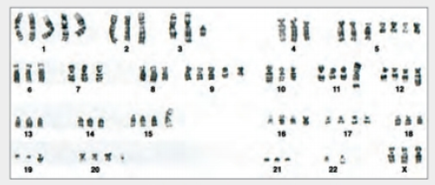
FIGURE 11.9 Karyotype of HeLa showing chromosomes in one cell.
Can you tell that this cell came from a female? How?

Trending nowThis is a popular solution!

Chapter 11 Solutions
Biology: The Unity and Diversity of Life (MindTap Course List)
Additional Science Textbook Solutions
Anatomy & Physiology
Microbiology: Principles and Explorations
Campbell Biology: Concepts & Connections (9th Edition)
Anatomy & Physiology: The Unity of Form and Function
Essentials of Human Anatomy & Physiology (11th Edition)
Becker's World of the Cell (9th Edition)


- HeLa Cells Are a Genetic Mess HeLa cells can vary in chromosome number. Defects in proteins that orchestrate cell division result in descendant cells with too many or too few chromosomes, an outcome that is one of the ha1lmarks of cancer cells. The panel of chromosomes in FIGURE 11.9, originally published in 1989, shows all of the chromosomes in a single metaphase HeLa cell. FIGURE 11.9 Karyotype of HeLa showing chromosomes in one cell. How many extra chromosomes does this cell have, compared to a normal human body cell?arrow_forwardHeLa Cells Are a Genetic Mess HeLa cells can vary in chromosome number. Defects in proteins that orchestrate cell division result in descendant cells with too many or too few chromosomes, an outcome that is one of the ha1lmarks of cancer cells. The panel of chromosomes in FIGURE 11.9, originally published in 1989, shows all of the chromosomes in a single metaphase HeLa cell. FIGURE 11.9 Karyotype of HeLa showing chromosomes in one cell. What is the chromosome number of this HeLa cell?arrow_forwardHeLa Cells Are a Genetic Mess HeLa cells can vary in chromosome number. Defects in proteins that orchestrate cell division result in descendant cells with too many or too few chromosomes, an outcome that is one of the ha1lmarks of cancer cells. The panel of chromosomes in FIGURE 11.9, originally published in 1989, shows all of the chromosomes in a single metaphase HeLa cell. FIGURE 11.9 Karyotype of HeLa showing chromosomes in one cell. How many extra chromosomes does this cell have, compared to a normal human body cell?arrow_forward
- After mitosis, each daughter cell contains genetic instructions that are ______ and _____ chromosome number of the parent cell. a. identical to the parent cells; the same b. identical to the parent cells; one-half the c. rearranged; the same d. rearranged; one-half thearrow_forwardHeLa Cells Are a Genetic Mess HeLa cells can vary in chromosome number. Defects in proteins that orchestrate cell division result in descendant cells with too many or too few chromosomes, an outcome that is one of the ha1lmarks of cancer cells. The panel of chromosomes in FIGURE 11.9, originally published in 1989, shows all of the chromosomes in a single metaphase HeLa cell. FIGURE 11.9 Karyotype of HeLa showing chromosomes in one cell. What is the chromosome number of this HeLa cell?arrow_forwardYOU DON'T NEED TO EXPLAIN THESE QUESTIONS JUST PROVIDE ANSWER DNA separates during ____ of mitosis. a)Prophase b)Telophase c)Metaphase d)Interphase e)Anaphase 2. What separates during mitosis? a)Single DNA strands b)Cytoplasm c)Sister chromatids d)Telomeres e)Homologous chromosomes 3. Oncogenes are associated with cancer because they Oncogenes are associated with cancer because they (select one answer) Cause cells to initiate a death pathway Fail to put a “brake” on the cell cycle Push cells through the cell cycle Divert cells into G0 phase Slow down the cell cyclearrow_forward
- The genes below have been knocked out (loss of function). Draw what the cell would look like during the appropriately affected stage of mitosis. State what stage you are depicting on your drawing. (Each gene knockout is occurring in a different cell; you should have a drawing of the affected cell for each). 1. Separase, 2. Cohesinarrow_forwardA person with cancer walks into the classroom. As a biology class, we ask if we can examine some of the cancerous cells and find that many of these cancerous cells have an extra chromosome 10 (trisomy) leading to more cyclin for forming the MPF being made, leading to uncontrollable cell division (i.e. tumor and progression to cancer). What would cause extra chromosomes in cells (be it somatic or gametic cells)? Select one: a. None of these choices are linked to how sister chromatids separate and would lead to extra chromosomes in daughter cells of cell division. b. APC not functioning correctly to add ubiquitin to the cohesins, thus sister chromatids do not split correctly during cell division c. MPF itself would cause more chromosome replication d. APC is overly functioning adding cohesins to the sister chromatidsarrow_forwardHuman body cells are diploid, which mean they______ . a. divide to form two cells b. have two full sets of chromosomes c. contain two chromosomesarrow_forward
- The microscope image above shows the human chromosomes from a white blood cell. To create the image, researchers put cells in culture under conditions that encourage the cells to divide. They bathed the cells in a hypotonic (low salt) solution, which caused the cells to swell until their plasma membrane burst open. They "squashed" the chromosomes to spread them out, and stained them with a dye to make them visible under the microscope. Human chromosomes are numbered from longest (1) to shortest (22) plus the sex chromosomes X and Y. In the image chromosome 1 is about 7 micrometers. Answer the following questions. 1) What word(s) in the description above indicates that the chromosomes are not from a cell undergoing meiosis? 2) Based on the size, shape and appearance of the chromosomes in the image, in what cell cycle stage was the cell that the chromosomes came from? How can you tell? 3) Does the image suggest that centromere sequences are always located in the middle of a…arrow_forward"Meiosis, oh meiosis, let me tell you what it means. It's a special kind of cell division with some funny little genes. It starts with a cell splitting in two, just like mitosis would; but from there, things get wild and wacky, and I'll tell you if I could. Instead of dividing once more, the cells each split again; creating four brand new cells, not just two like back then. And here's where the magic happens, with chromosomes all pairing up, then swapping bits of info, a dizzying process that can't be summed up. When all is said and done, those four new cells are not quite the same; they've got half the number of chromosomes, and a whole new genetic game. Meiosis, oh meiosis, it's a puzzle that's so much fun. And if you study it like I have, you'll see how it all gets done!"arrow_forwardFigure 6.4 Which of the following is the correct order of events in mitosis?a. Sister chromatids line up at the metaphase plate. The kinetochore becomes attached to the mitotic spindle. The nucleus re-forms and the cell divides. The sister chromatids separate.b. The kinetochore becomes attached to the mitotic spindle. The sister chromatids separate. Sister chromatids line up at the metaphase plate. The nucleus re-forms and the cell divides.c. The kinetochore becomes attached to metaphase plate. Sister chromatids line up at the metaphase plate. The kinetochore breaks down and the sister chromatids separate. The nucleus re-forms and the cell divides.d. The kinetochore becomes attached to the mitotic spindle. Sister chromatids line up at the metaphase plate. The kinetochore breaks apart and the sister chromatids separate. The nucleus re-forms and the cell divides.arrow_forward
 Biology: The Unity and Diversity of Life (MindTap...BiologyISBN:9781337408332Author:Cecie Starr, Ralph Taggart, Christine Evers, Lisa StarrPublisher:Cengage Learning
 Biology: The Unity and Diversity of Life (MindTap...BiologyISBN:9781305073951Author:Cecie Starr, Ralph Taggart, Christine Evers, Lisa StarrPublisher:Cengage Learning
 Biology Today and Tomorrow without Physiology (Mi...BiologyISBN:9781305117396Author:Cecie Starr, Christine Evers, Lisa StarrPublisher:Cengage Learning
 Human Biology (MindTap Course List)BiologyISBN:9781305112100Author:Cecie Starr, Beverly McMillanPublisher:Cengage Learning