Concept explainers
To review:
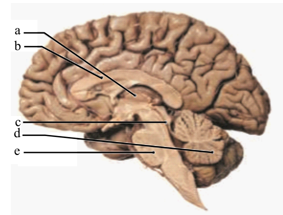
Label the following terms in the given diagram of the midsagittal section of the brain: pons, corpus callosum, cerebellum, thalamus, and cerebral aqueduct.
Introduction:
The brain is the prime organ of the CNS (central nervous system). It is divided into six major regions on the basis of structure and function, which are cerebrum, cerebellum, medulla oblongata, pons, diencephalon, and mesencephalon.
Explanation of Solution
The structures of the midsagittal section of the brain are labeled as below:
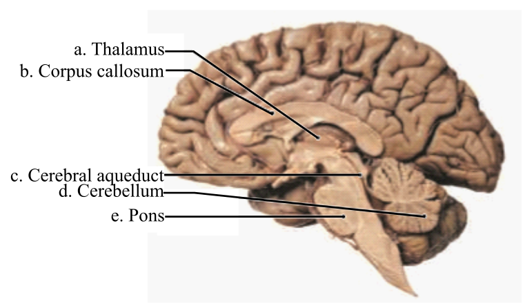
Thalamus: It is a part of the diencephalon that is further divided into right and left thalamus. This region is assigned the duty to relay and process sensory information across the brain.
Corpus callosum: It is a bundle of commissural fibers that connects the two cerebral hemispheres. It functions to transfer information from one hemisphere to another. It contains the largest portion of the white matter in the brain.
Cerebral aqueduct: It is a part of the mesencephalon or midbrain. It connects the third and fourth ventricles within the diencephalon and mesencephalon respectively. It is filled with cerebrospinal fluid (CSF), which flows into the ventricles.
Cerebellum: It is a part of the hindbrain and called as the small brain. It functions to relay motor sensations and is involved in intellectual development. It also maintains the posture of the body.
Pons: It is a part of the brainstem and is located below the midbrain, above the medulla oblongata and anterior to the cerebellum. It functions to relay sensory information between the thalamus and the cerebellum. It is responsible for the maintenance of body posture, hearing, taste, touch, pain, facial expression, chewing, salivation, and secretion of tears.
Therefore, the structures of a midsagittal section of the brain are:
| a | Thalamus |
| b | Corpus callosum |
| c | Cerebral aqueduct |
| d | Cerebellum |
| e | Pons |
Want to see more full solutions like this?
Chapter 16 Solutions
Human Anatomy, Books a la Carte Plus Mastering A&P with Pearson eText - Access Card Package (9th Edition)


- Julio D., who had recently retired, was enjoying an afternoon of playing golf when suddenly he experienced a severe headache and dizziness. These symptoms were quickly followed by numbness and partial paralysis on the tipper right side of his body, accompanied by an inability to speak. After being rushed to the emergency room, Julio was diagnosed as having suffered a stroke. Given the observed neurological impairment, what areas of his brain were affected?arrow_forwardWatch this video (http://openstaxcollege.org/l/lumbarpuncture) that describes the procedure known as the lumbar puncture, a medical procedure used to sample the CSF. Because of the anatomy of the CNS, it is a relative safe location to insert a needle. Why is the lumbar puncture performed in the lower lumbar area of the vertebral column?arrow_forwardWatch this animation (http://openstaxcollege.org/l/CSFflow) that shows the flow of CSF through the brain and spinal cord, and how it originates from the ventricles and then spreads into the space within the meninges, where the fluids then move into the venous sinuses to return to the cardiovascular circulation. What are the structures that produce CSF and where are they found? How are the structures indicated in this animation?arrow_forward

 Fundamentals of Sectional Anatomy: An Imaging App...BiologyISBN:9781133960867Author:Denise L. LazoPublisher:Cengage Learning
 Anatomy & PhysiologyBiologyISBN:9781938168130Author:Kelly A. Young, James A. Wise, Peter DeSaix, Dean H. Kruse, Brandon Poe, Eddie Johnson, Jody E. Johnson, Oksana Korol, J. Gordon Betts, Mark WomblePublisher:OpenStax College
 Human Physiology: From Cells to Systems (MindTap ...BiologyISBN:9781285866932Author:Lauralee SherwoodPublisher:Cengage Learning