Videos
Building Better Bones Tiffany, shown in FIGURE 35.22, was born with multiple fractures in her arms and legs. By age six, she had undergone surgery to correct more than 200 bone fractures. Her fragile, easily broken bones are symptoms of osteogenesis imperfecta (OI), a genetic disorder caused by a mutation in a gene for collagen. As bones develop, collagen forms a scaffold for deposition of mineralized bone tissue. The scaffold forms improperly in children with OI. FIGURE 35.22 also shows the results of a test of a new drug. Treated children, all less than two years old, were compared to similarly affected children of the same age who were not treated with the drug.
| Vertebral | |||
| Treated | area in cm2 | Fractures | |
| child | (Initial) | (Final) | per year |
| 1 | 14.7 | 16.7 | 1 |
| 2 | 15.5 | 16.9 | 1 |
| 3 | 6.7 | 16.5 | 6 |
| 4 | 7.3 | 11.8 | 0 |
| 5 | 13.6 | 14.6 | 6 |
| 6 | 9.3 | 15.6 | 1 |
| 7 | 15.3 | 15.9 | 0 |
| 8 | 9.9 | 13.0 | 4 |
| 9 | 10.5 | 13.4 | 4 |
| Mean | 11.4 | 14.9 | 2.6 |
| Vertebral | |||
| Treated | area in cm2 | Fractures | |
| child | (Initial) | (Final) | per year |
| 1 | 18.2 | 13.7 | 4 |
| 2 | 16.5 | 12.9 | 7 |
| 3 | 16.4 | 11.3 | 8 |
| 4 | 13.5 | 7.7 | 5 |
| 5 | 16.2 | 16.1 | 8 |
| 6 | 18.9 | 17.0 | 6 |
| Mean | 16.6 | 13.1 | 6.3 |
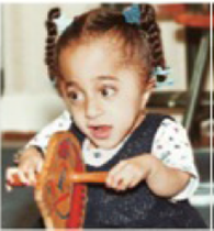
FIGURE 35.22 Results of a clinical trial of a drug treatment for osteogenesis imperfecta (OI), which affects the child shown at right. Nine children with OI received the drug. Six others were untreated controls. Surface area of certain vertebrae was measured before and after treatment. Fractures occurring during the 12 months of the trial were also recorded.
3. How did the rate of fractures in the two groups compare?
Want to see the full answer?
Check out a sample textbook solution
Chapter 35 Solutions
Biology: The Unity and Diversity of Life (MindTap Course List)


- Building Better Bones Tiffany, shown in FIGURE 35.22, was born with multiple fractures in her arms and legs. By age six, she had undergone surgery to correct more than 200 bone fractures. Her fragile, easily broken bones are symptoms of osteogenesis imperfecta (OI), a genetic disorder caused by a mutation in a gene for collagen. As bones develop, collagen forms a scaffold for deposition of mineralized bone tissue. The scaffold forms improperly in children with OI. FIGURE 35.22 also shows the results of a test of a new drug. Treated children, all less than two years old, were compared to similarly affected children of the same age who were not treated with the drug. Vertebral Treated area in cm2 Fractures child (Initial) (Final) per year 1 14.7 16.7 1 2 15.5 16.9 1 3 6.7 16.5 6 4 7.3 11.8 0 5 13.6 14.6 6 6 9.3 15.6 1 7 15.3 15.9 0 8 9.9 13.0 4 9 10.5 13.4 4 Mean 11.4 14.9 2.6 Vertebral Treated area in cm2 Fractures child (Initial) (Final) per year 1 18.2 13.7 4 2 16.5 12.9 7 3 16.4 11.3 8 4 13.5 7.7 5 5 16.2 16.1 8 6 18.9 17.0 6 Mean 16.6 13.1 6.3 FIGURE 35.22 Results of a clinical trial of a drug treatment for osteogenesis imperfecta (OI), which affects the child shown at right. Nine children with OI received the drug. Six others were untreated controls. Surface area of certain vertebrae was measured before and after treatment. Fractures occurring during the 12 months of the trial were also recorded. 4. Do these results shown support the hypothesis that this drug, which slows bone breakdown, can increase bone growth and reduce fractures in young children with OI?arrow_forwardBuilding Better Bones Tiffany, shown in FIGURE 35.22, was born with multiple fractures in her arms and legs. By age six, she had undergone surgery to correct more than 200 bone fractures. Her fragile, easily broken bones are symptoms of osteogenesis imperfecta (OI), a genetic disorder caused by a mutation in a gene for collagen. As bones develop, collagen forms a scaffold for deposition of mineralized bone tissue. The scaffold forms improperly in children with OI. FIGURE 35.22 also shows the results of a test of a new drug. Treated children, all less than two years old, were compared to similarly affected children of the same age who were not treated with the drug. Vertebral Treated area in cm2 Fractures child (Initial) (Final) per year 1 14.7 16.7 1 2 15.5 16.9 1 3 6.7 16.5 6 4 7.3 11.8 0 5 13.6 14.6 6 6 9.3 15.6 1 7 15.3 15.9 0 8 9.9 13.0 4 9 10.5 13.4 4 Mean 11.4 14.9 2.6 Vertebral Treated area in cm2 Fractures child (Initial) (Final) per year 1 18.2 13.7 4 2 16.5 12.9 7 3 16.4 11.3 8 4 13.5 7.7 5 5 16.2 16.1 8 6 18.9 17.0 6 Mean 16.6 13.1 6.3 FIGURE 35.22 Results of a clinical trial of a drug treatment for osteogenesis imperfecta (OI), which affects the child shown at right. Nine children with OI received the drug. Six others were untreated controls. Surface area of certain vertebrae was measured before and after treatment. Fractures occurring during the 12 months of the trial were also recorded. 2. How many of the untreated children showed an increase in vertebral area?arrow_forwardBuilding Better Bones Tiffany, shown in FIGURE 35.22, was born with multiple fractures in her arms and legs. By age six, she had undergone surgery to correct more than 200 bone fractures. Her fragile, easily broken bones are symptoms of osteogenesis imperfecta (OI), a genetic disorder caused by a mutation in a gene for collagen. As bones develop, collagen forms a scaffold for deposition of mineralized bone tissue. The scaffold forms improperly in children with OI. FIGURE 35.22 also shows the results of a test of a new drug. Treated children, all less than two years old, were compared to similarly affected children of the same age who were not treated with the drug. Vertebral Treated area in cm2 Fractures child (Initial) (Final) per year 1 14.7 16.7 1 2 15.5 16.9 1 3 6.7 16.5 6 4 7.3 11.8 0 5 13.6 14.6 6 6 9.3 15.6 1 7 15.3 15.9 0 8 9.9 13.0 4 9 10.5 13.4 4 Mean 11.4 14.9 2.6 Vertebral Treated area in cm2 Fractures child (Initial) (Final) per year 1 18.2 13.7 4 2 16.5 12.9 7 3 16.4 11.3 8 4 13.5 7.7 5 5 16.2 16.1 8 6 18.9 17.0 6 Mean 16.6 13.1 6.3 FIGURE 35.22 Results of a clinical trial of a drug treatment for osteogenesis imperfecta (OI), which affects the child shown at right. Nine children with OI received the drug. Six others were untreated controls. Surface area of certain vertebrae was measured before and after treatment. Fractures occurring during the 12 months of the trial were also recorded. 1. An increase in vertebral area during the 12-month period of the study indicates bone growth. How many of the treated children showed such an increase?arrow_forward
- The cell found in bone that makes the bone is called an. osteoblast osteocyte osteoclast osteonarrow_forwardThe cells responsible for bone resorption are ___________ . osteoclasts osteoblasts fibroblasts osteocytesarrow_forwardWhy is cartilage slow to heal? because it eventually develops into bone because it is semi-solid and flexible because it does not have a blood supply because endochondral ossification replaces all cartilage with bonearrow_forward
- Question:- Which of the following is true regarding endochondrial ossification? a. Committed mesenchymal cells differentiate into cartilage cells and condense into compact nodules b. Apoptosis of undifferentiated mesenchyme allows for blood vessel integration c. Differentiated chondrocytes begin apoptosis and form the blood vessels d. Hypertrophic chondrocytes begin to compact into nodules at primary ossification centerarrow_forwardA race between two runners is often used as an analogy to describe the mechanism of endochondral ossification. Review endochondral ossification. In endochondral ossification, who are the two runners? Did each runner begin the race at the same time? Who is in the lead? How does the race end? I have some ideas but cannot decide. I was first thinking condrocytes and osteoblasts with osteoblasts finishing first with the compact bone, but then I was thinking of blasts and clasts as one builds and one removes. Also, I thought, well maybe, cartilage and osteoblasts.arrow_forwardWhy are osteocytes spread out in bone tissue? They develop from mesenchymal cells. They are surrounded by osteoid. They travel through the capillaries. Formation of osteoid spreads out the osteoblasts that fanned the ossification centers.arrow_forward
- The epiphyseal plate: is arranged as rods or plates contains the bone’s blood vessels and nerve fibers is responsible for the lengthwise growth of long bones synthesizes and secretes bone matrixarrow_forwardThe cell found in bone that breaks it down is called an. osteoblast osteocyte osteoclast osteonarrow_forwardThe Haversian canal: is arranged as rods or plates contains the bone’s blood vessels and nerve fibers is responsible for the lengthwise growth of long bones synthesizes and secretes matrixarrow_forward
 Biology: The Unity and Diversity of Life (MindTap...BiologyISBN:9781337408332Author:Cecie Starr, Ralph Taggart, Christine Evers, Lisa StarrPublisher:Cengage Learning
 Biology: The Unity and Diversity of Life (MindTap...BiologyISBN:9781305073951Author:Cecie Starr, Ralph Taggart, Christine Evers, Lisa StarrPublisher:Cengage Learning
 Anatomy & PhysiologyBiologyISBN:9781938168130Author:Kelly A. Young, James A. Wise, Peter DeSaix, Dean H. Kruse, Brandon Poe, Eddie Johnson, Jody E. Johnson, Oksana Korol, J. Gordon Betts, Mark WomblePublisher:OpenStax College
 Human Biology (MindTap Course List)BiologyISBN:9781305112100Author:Cecie Starr, Beverly McMillanPublisher:Cengage Learning
 Biology: The Dynamic Science (MindTap Course List)BiologyISBN:9781305389892Author:Peter J. Russell, Paul E. Hertz, Beverly McMillanPublisher:Cengage Learning
 Biology 2eBiologyISBN:9781947172517Author:Matthew Douglas, Jung Choi, Mary Ann ClarkPublisher:OpenStax