
Videos
To label: The urinary structures given in the figure.
Introduction: The urinary system is the drainage system of the body for removing wastes in the form of urine. For this process, the parts of the urinary system should work together in coordination. The mammalian urinary system includes kidneys, ureters, urinary bladder, and urethra.
Explanation of Solution
Pictorial representation: The organs of the female urinary system are given in the Fig.1.
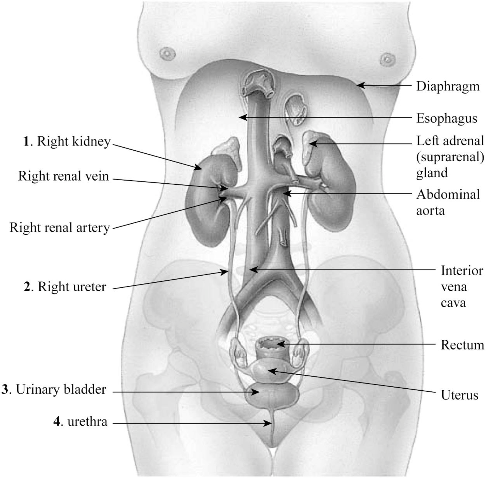
Fig.1: The organs of the female urinary system.
1. Right kidney: The kidney is an important organ of the excretory system. The right kidney is slightly longer than the left kidney.
2. Right ureter: The right ureter is located on the right side of the abdomen and is a long fibromuscular tube that transports urine from the right kidney to the urinary bladder. Urine makes the renal pelvis to contract in rhythm to drive urine through the ureters into the urinary bladder.
3. Urinary bladder: The wall of the urinary bladder has the detrusor muscle; it allows the storage of urine and contraction during urination and release of urine.
4. Urethra: Urethra is a tube that carries urine to the outside of the body. The bladder is joined to the urethra through the bladder neck that consists of internal sphincter muscles.
Pictorial representation: The location and coverings of the kidneys are given in Fig.2.
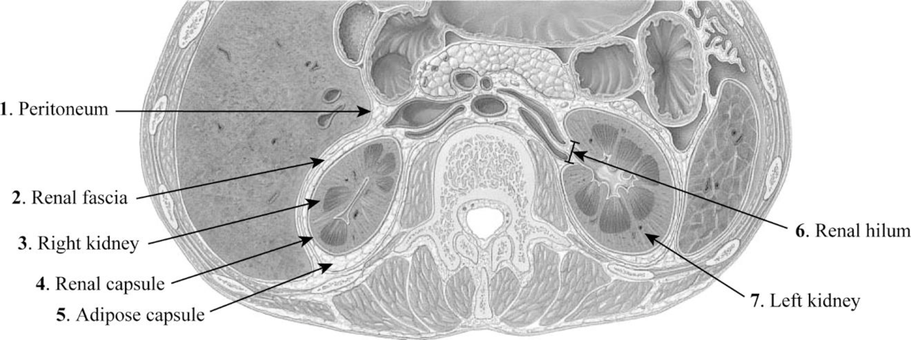
Fig.2: The location and coverings of the kidneys.
1. Peritoneum: The peritoneum is a sheet-like membrane that forms two layers and a space called the peritoneal cavity.
2. Renal fascia: The renal fascia is the layer of irregular connective tissue. This layer covers the adipose capsule and encapsulates the kidney. It passes anteriorly to the kidney, abdominal aorta, and renal vessels and posteriorly connects to the psoas fascia.
3. Right kidney: The right kidney is located on the right side of the abdomen and is connected to the right ureters through which urine reaches the urinary bladder.
4. Renal capsule: The fibrous capsule of the kidney is also known as the renal capsule. It is directly adhered to the external surface of the kidney.
5. Adipose capsule: The adipose capsule is the structure of the kidney that is located between the renal fascia and renal capsule. It is the perirenal fat that covers the renal capsule. This layer provides protection to the kidney from damage.
6. Renal hilum: The renal hilum is a depression in the surface of the kidney where blood vessels and nerves enter and exit.
7. Left kidney: The left kidney is located on the left side of the abdomen and is slightly shorter than the right kidney.
Pictorial representation: The internal structure of the kidney is given in Fig.3.
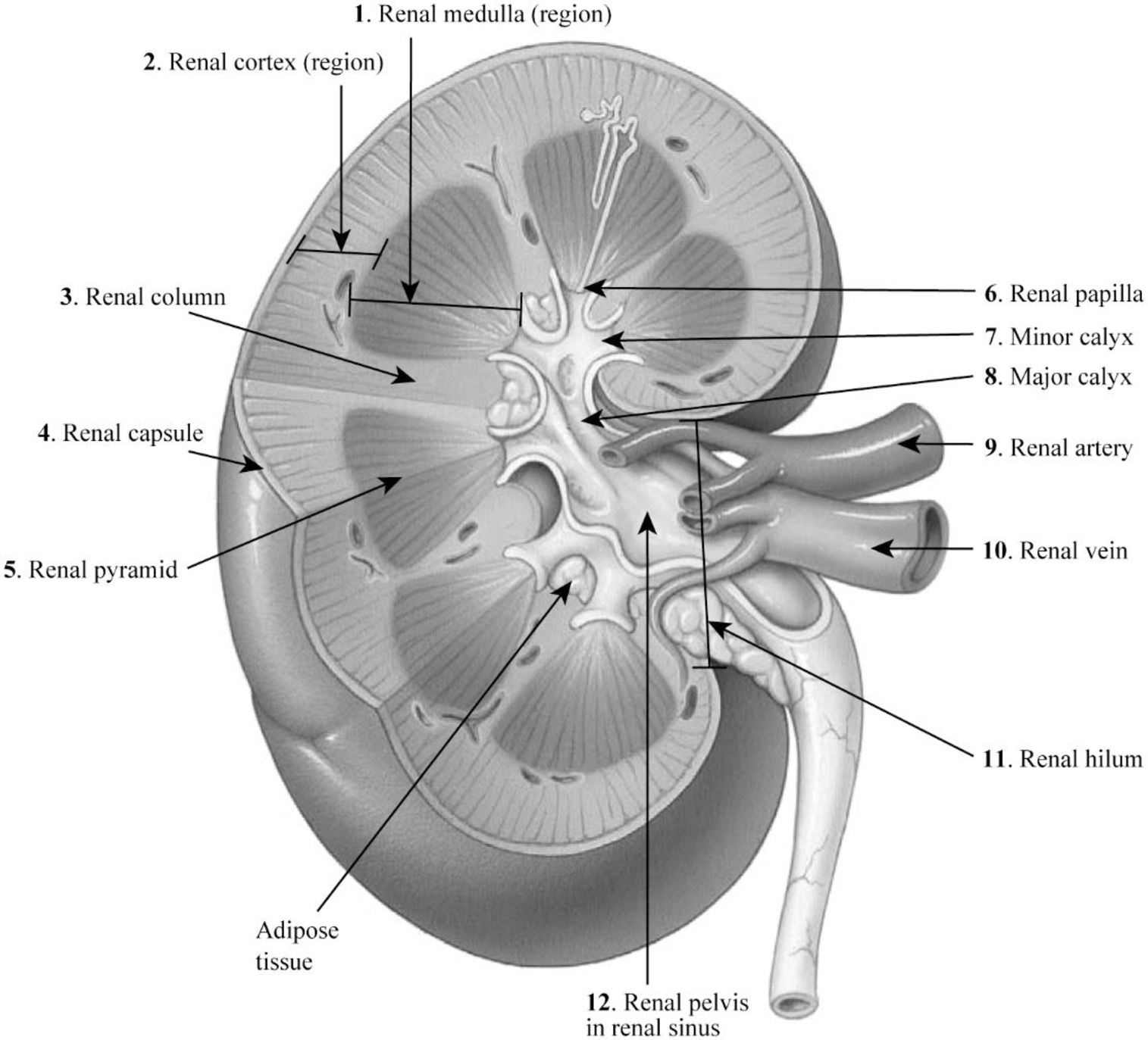
Fig.3: The internal structure of the kidney.
1. Renal medulla: The renal medulla is the deep region to the cortex of the kidney that contains 8–12 renal pyramids consisting of collecting ducts.
2. Renal cortex: The renal cortex is the outer portion of the kidney. It is a smooth outer zone present between the renal capsule and the renal medulla.
3. Renal column: The medullary renal cortex extensions present in between renal pyramids are called the renal column. This allows the renal cortex to be better attached, and each column consists of blood vessel and urinary tube lines.
4. Renal capsule: The renal capsule is composed of dense irregular connective tissue and maintains the kidney’s shape, protects from trauma, and prevents infectious pathogens from penetrating the kidney.
5. Renal pyramid: The renal pyramid is a cone-shaped tissue that consists of various tubules that transport located within the renal medulla. At the renal papilla, the renal pyramids drain urine into the minor calyx in the kidney.
6. Renal papilla: The renal papilla is the peak of the renal pyramid. At the renal papilla, the renal pyramids drain urine into the minor calyx in the kidney.
7. Minor calyx: The renal pelvis connects all the renal tubules and forms a cup-like region called calyces. The minor calyces surround the peak of the renal pyramids.
8. Major calyx: Two to three minor calyces unite to form the major calyx. Urine enters the papillary duct that is located within the renal papilla and then passes through the spaces (minor calyx, major calyx, and renal pelvis) within the renal sinus of the kidney.
9. Renal artery: The renal arteries carry a very large amount of blood from the heart to the kidneys for the process of filtration.
10. Renal vein: A large amount of blood enters the kidneys at the hilum through the renal artery and leaves the kidneys through the renal vein.
11. Renal hilum: A large amount of blood enters the kidneys at the hilum through the renal artery and leaves the kidneys through the renal vein.
12. Renal pelvis in renal sinus: The renal pelvis in the renal sinus connects all the renal tubules and forms a cup-like region called calyces. The minor calyces surround the peak of the renal pyramids. At the renal papilla, the renal pyramids drain urine into the minor calyx in the kidney.
Want to see more full solutions like this?
Chapter 36 Solutions
Laboratory Manual for Anatomy and Physiology, 6e Loose-Leaf Print Companion


 Human Anatomy & Physiology (11th Edition)BiologyISBN:9780134580999Author:Elaine N. Marieb, Katja N. HoehnPublisher:PEARSON
 Biology 2eBiologyISBN:9781947172517Author:Matthew Douglas, Jung Choi, Mary Ann ClarkPublisher:OpenStax
 Anatomy & PhysiologyBiologyISBN:9781259398629Author:McKinley, Michael P., O'loughlin, Valerie Dean, Bidle, Theresa StouterPublisher:Mcgraw Hill Education,
 Molecular Biology of the Cell (Sixth Edition)BiologyISBN:9780815344322Author:Bruce Alberts, Alexander D. Johnson, Julian Lewis, David Morgan, Martin Raff, Keith Roberts, Peter WalterPublisher:W. W. Norton & Company
 Laboratory Manual For Human Anatomy & PhysiologyBiologyISBN:9781260159363Author:Martin, Terry R., Prentice-craver, CynthiaPublisher:McGraw-Hill Publishing Co.
 Inquiry Into Life (16th Edition)BiologyISBN:9781260231700Author:Sylvia S. Mader, Michael WindelspechtPublisher:McGraw Hill Education