
Concept explainers
Videos
If you compare electron micrographs of a relaxed skeletal muscle fiber and a fully contracted muscle fiber, which would you see only in the relaxed fiber?
a. Z discs
b. Sarcomeres
c. I bands
d. A bands
e. H zones
Introduction:
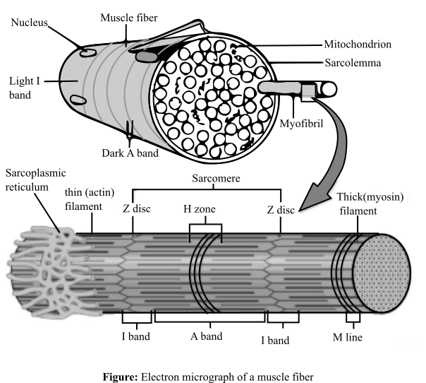
The skeletal muscle fibers are made of striated muscle tissues, which consists of alternating light (I) and dark bands (A). I-band consists of a dark region known as Z disc. On the other hand, A band consists of a light region known as H-zone. I-band consists only of thin actin filaments, while the A-band consists of both actin and myosin filaments. All these units of a muscle fiber makes sarcomere. The micrograph of a sarcomere is shown as below:
Answer to Problem 1MC
Correct answer:
I bands and the H zones of the skeletal muscle fiber will be visible only in the micrograph of the relaxed fiber.
Explanation of Solution
Explanation for the correct answer:
Option (c) and option (e) are given as I bands, and H zones, respectively. A sarcomere is basically a unit of the striated muscle tissue. Repeating units of Z lines are present in this and the actin molecules are bounded to these lines forming a border in the sarcomere. Alternating I which are light and A which are dark bands are present. When looking closer, I band comprises of a midline interruption which is known as Z disc while in the A band there is a lighter middle section known as H zone. During the muscle contraction, the actin filaments move over the myosin filaments according to the sliding filament theory. I-band and the H zone tends to overlap when a muscle is contracted. When they overlap it becomes difficult to observe I and H bands. Therefore, these will only be visible in the relaxed position. Hence, option (c) and option (e) are correct.
Explanation for the incorrect answers:
Option (a) is given as Z discs. The Z discs are dark stripes, which marks the ending of a sarcomere. These dark zones can be clearly seen in both the contracted and relaxed electron micrographs of the skeletal muscles. So, it is an incorrect option.
Option (b) is given as sarcomere. This is the part of the muscle composing of the thin and thick filaments. These thick and thin filaments forming the sarcomere are the basic unit of the muscle tissue present between the two Z discs. Therefore, sarcomere will be visible in both the contracted as well as a relaxed muscle fiber. So, it is an incorrect option.
Option (d) is given as A bands. These are the darker areas of the sarcomere. These bands do not shrink, when the muscle contracts, and hence, can be seen both in relaxed and contracted position. So, it is an incorrect option.
Hence, options (a), (b), and (d) are incorrect.
Thus, in a relaxed state, I-band and H-zone will be visible, which will disappear during contraction of the muscle.
Want to see more full solutions like this?
Chapter 6 Solutions
ESSENTIALS OF HUMAN ANATOMY & PHYSIOLOG


- Which component is responsible for initially stimulating a muscle contraction? a. proteins b. electrochemical signals c. plasma membranes d. striationsarrow_forwardIn muscle cells, magnesium ions compete with calcium ions for binding sites on troponin molecules. If a person has too high a concentration of magnesium ions in the blood, magnesium ions can prevent calcium ions from binding troponin. A) What effect would this have on muscle contraction (strengthen, weaken, or no effect)? B) Use your knowledge of how muscle fibers contract to explain your answer in part A.arrow_forwardA skeletal muscle motor unit A.Is the number of muscle cells innervated by one motor neuron. B.Is the number of motor neurons innervating one muscle cell. C.Size determines the ability to control a muscle D.Size determines how much a muscle contracts at any one time.arrow_forward
- In a skeletal muscle fiber, Ca2+ is released from a. ACh receptors. b. the motor end plate. c. the sarcoplasmic reticulum. d. the sarcolemma and T-tubules.arrow_forwardIf a muscle cell had very short T-tubules, how much tension would the muscle fiber create, relative to a normal muscle fiber? Assume sarcoplasmic reticulum can still react to activity at the neuromuscular junction. a)Less tension would be created. b) No difference in tension creation. c) More tension would be created. d) No tension would be created.arrow_forwardWhen sarcomeres contract during muscle contraction, which of the following occurs? A. The myosin filaments lengthen. B. The myosin filaments "walk" along the actin microfilaments. C. The myosin filaments shorten. D. The actin filaments shorten.arrow_forward
- Outline the role of calcium ions on muscle contraction during a biceps curl. In your answer: A) Describe the role of the nervous system in stimulating the release of calcium ions. b) Outline the role calcium ions play in the 'sliding filament theory?'arrow_forwardA bacterial toxin is known to block the release of ACh at the motor end plate of skeletal muscle. Consequently, a. the skeletal muscle contracts with increasing force. b. the skeletal muscle contracts with increasing frequency. c. the ability to stimulate the muscle is impaired. d. other neurotransmitters would stimulate the muscle.arrow_forwarda. Describe the relationship between stimulus voltage and the force of contraction b. What was the smallest voltage required to produce a contraction (the threshold voltage)? What proportion of the fibers in the muscle do you think were contracting to produce this small response? c. What do you conclude happened to the number of fibers contracting as the voltage was raised from threshold to that required to produce a maximal contraction?arrow_forward
- Which statement about striated skeletal muscle is true? A. The tension generated by a muscle is invariable. B. Mechanical summation of twitches in a muscle fiber leads to a graded increase in the tension that is above that generated by a single twitch. C. A single action potential arriving at the neuromuscular junction is not sufficient to cause a muscle fiber to twitch. D. Muscle twitches are able to mechanically sum when Ca2+ is quickly and completely removed from the sarcoplasm between action potentials. E. An action potential in the muscle cell activates contraction by releasing Na+ into the sarcoplasm.arrow_forwardWhat happens to sarcomeres when actin and myosin filaments in a muscle fiber interact during the contraction phase? A) sarcomeres usually fatigue B) sarcomeres usually get shorter C) sarcomeres usually get longer D) sarcomeres stay the samearrow_forwardWhich of the following statements best describes the sliding filament mechanism of muscle contraction? a. Actin and myosin filaments do not shorten, but rather, slide past each other. b. Actin and myosin filaments shorten and slide past each other. c. As they slide past each other, actin filaments shorten, but myosin filaments do not shorten. d. As they slide past each other, myosin filaments shorten, but actin filaments do not shorten.arrow_forward
 Concepts of BiologyBiologyISBN:9781938168116Author:Samantha Fowler, Rebecca Roush, James WisePublisher:OpenStax College
 Human Physiology: From Cells to Systems (MindTap ...BiologyISBN:9781285866932Author:Lauralee SherwoodPublisher:Cengage Learning
 Comprehensive Medical Assisting: Administrative a...NursingISBN:9781305964792Author:Wilburta Q. Lindh, Carol D. Tamparo, Barbara M. Dahl, Julie Morris, Cindy CorreaPublisher:Cengage Learning
 Human Biology (MindTap Course List)BiologyISBN:9781305112100Author:Cecie Starr, Beverly McMillanPublisher:Cengage Learning