
Concept explainers
Videos
RIPs as Cancer Drugs Researchers are taking a page from the structure-function relationship of RIPs in their quest for cancer treatments. The most toxic RIPs, remember, have one domain that interferes with ribosomes, and another that carries them into cells. Melissa Cheung and her colleagues incorporated a peptide that binds to skin cancer cells into the enzymatic part of an RIP, the E. coli Shiga-like toxin. The researchers created a new RIP that specifically kills .skin cancer cells, which are notoriously resistant to established therapies. Some of their results are shown in FIGURE 9.17.
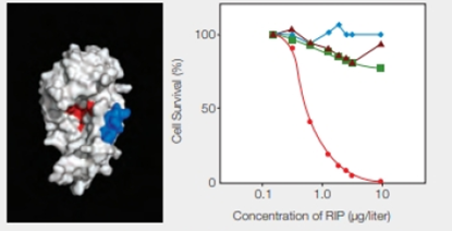
FIGURE 9.17 Effect of an engineered RIP on cancer cells. The model on the left shows the enzyme portion of E. coli Shiga-like toxin engineered to carry a small sequence of amino acids (in blue) that targets skin cancer cells. (Red indicates the active site.) The graph on the right shows the effect of this engineered RIP on human cancer cells of the skin ( ); breast (
 ) liver (
 ); and prostate (
 ).
).
Which cells had the greatest response to an increase in concentration of the engineered RIP?
To determine: The type of cells that had the greatest response to an increase in the concentration of the engineered RIP.
Introduction: Ribosome-inactivating proteins (RIPs) inactivate the ribosomes and prevent protein synthesis in a cell. The toxic RIPs have a domain that makes them enter into the cell and another domain that interferes with the ribosome. They have antiviral and anticancer properties and are used to design drugs for HIV and cancer.
Answer to Problem 1DAA
Correct answer: The greatest response in the form of fall in cell’s survival percentage with an increase in the concentration of engineered RIP is seen in the skin cancer cells.
Explanation of Solution
As given in the problem statement, Researcher M and her colleagues incorporated a peptide into the enzymatic part of a RIP, the E. coli Shiga-like toxin. The peptide specifically binds to the skin cancer cells, and thus, the newly synthesized RIP kills the skin cancer cells.
Refer Fig. 9.17, “Effect of an engineered RIP on cancer cells”, in the textbook. The model shown on the left indicates a blue-colored enzyme region of E. coli Shiga-like toxin that is engineered to carry the peptide sequence specific for the skin cancer cells. The red color indicates the active site of RIP.
The graphical representation that is shown in Fig. 9.17 on the right side indicates the effect of the engineered RIP on different human cancer cells indicated by different colors and shapes. They include skin, breast, liver, and prostate cancer cells with red, blue, brown, and green color, respectively. The concentration of RIP (µg/liter) is plotted with the percentage of cell survival. As shown in the graph, as the concentration of RIP increases, there is a significant drop in the skin cancer cells percentage. It reaches to zero at RIP concentration of 10 µg/liter. In the case of the other cancer cells, there is lesser variability.
Thus, the greatest response in the form of fall in cell’s survival percentage with an increase in the concentration of engineered RIP is seen in the skin cancer cells.
Want to see more full solutions like this?
Chapter 9 Solutions
Biology: The Unity and Diversity of Life (MindTap Course List)
Additional Science Textbook Solutions
Microbiology: An Introduction
Campbell Essential Biology with Physiology (5th Edition)
Biological Science
Campbell Essential Biology (6th Edition) - standalone book
Human Anatomy & Physiology (11th Edition)


- Polypeptide folding is often mediated by other proteins called chaperones. Describe how a mutant chaperone protein might be responsible for a genetic disorder involving an enzyme.arrow_forwardWhich model (2D topology or trRosetta) is likely a better representation of the actual protein AlaE, why?arrow_forwardLoop regions play important roles in the secondary structure of protein. Define loop region and give three (3) of the rolesarrow_forward
- Which of the following statements are correct about the role of chaperones in protein folding (select all that appy)? A. Chaperones accelerate the rate of protein folding B. Presence of chaperones contradicts dogma that structure of protein is only determined by amino acid sequence C. Chapreone promotion of native folding is an ATP-dependent process D. Chaperones work by stabilizing hydrophobic patches on proteins E. A cell line with the GroEL and GroES deleted would accumulate more unfolded proteinarrow_forwardAnfinsen studied protein folding using RNaseA. In one experiment we didn’t discuss in class, he heated RNase in the presence of BME, then he transferred the protein out of water into benzene, and cooled the protein in the presence of O2. Under these conditions, did Anfinsen find the protein was active? Justify your answer.arrow_forwardWhat forces come into play with protein folding (please explain this at the molecular level)?arrow_forward
- Which factor has NOT been shown to play a role in determining the specificity of protein kinases? a. protein tertiary structure b. protein quaternary structure c. primary sequence at phosphorylation site d. disulfide bonds near the phosphorylation site e. residues near the phosphorylation sitearrow_forwardProtein Structure and Function A common strategy in the regulation of protein function is to alter its structure. Describe two specific strategies used by the cell to alter a protein’s structure, thereby altering its function.arrow_forwardIn the early days of research on protein synthesis, some scientists observed that their most highly purified ribosome preparations, containing almost exclusively single ribosomes, were less active than preparations that were less highly purified. Suggest an explanation for this observation.arrow_forward
- SCIENTIFIC INQUIRY Knowing that the genetic code is almostuniversal, a scientist uses molecular biological methods toinsert the human β-globin gene (shown in Figure 17.12) intobacterial cells, hoping the cells will express it and synthesizefunctional β-globin protein. Instead, the protein produced isnonfunctional and is found to contain many fewer amino acidsthan does β-globin made by a eukaryotic cell. Explain whyarrow_forwardChoose if it's true or false Different from RNA since in the latter the internucleotide linkages are between C-2’ and C-5’ A long chain polymer in which the internucleotide linkages are of the diester type between C-3’ and C-5’ Usually present in tissues as a nucleoprotein and cannot be separated from its protein component Hydrolyzed by weak alkali (pH 9 to 100°C)arrow_forwardTo better understand the effects of palmitoylation on protein X, researchers want to chemically attach a palmitic acid at a specific position in this protein. The researchers first try to feed cells with palmitic acid or an amino acid with a C16 side chain. Neither of the initial experiments is successful in helping the researchers learn more about the biological role of palmitoylated protein X. Why? Suggest a strategy to specifically incorporate a palmitoylation site into protein X.arrow_forward
 Biology: The Unity and Diversity of Life (MindTap...BiologyISBN:9781305073951Author:Cecie Starr, Ralph Taggart, Christine Evers, Lisa StarrPublisher:Cengage Learning
 Human Heredity: Principles and Issues (MindTap Co...BiologyISBN:9781305251052Author:Michael CummingsPublisher:Cengage Learning
 BiochemistryBiochemistryISBN:9781305577206Author:Reginald H. Garrett, Charles M. GrishamPublisher:Cengage Learning