(a) A medical researcher is using the uniform electric field (E = 2400 NC-¹) generated by an electrophoresis setup to isolate a charged protein of interest (q = +160e) from an uncharged patient sample. As shown in panel (A), when the patient sample is loaded between the positive and negative terminal, the charged protein of interest migrates 12 cm toward the negative terminal.
(a) A medical researcher is using the uniform electric field (E = 2400 NC-¹) generated by an electrophoresis setup to isolate a charged protein of interest (q = +160e) from an uncharged patient sample. As shown in panel (A), when the patient sample is loaded between the positive and negative terminal, the charged protein of interest migrates 12 cm toward the negative terminal.

Related questions
Question
Transcribed Image Text:Question 5 Electromagnetic phenomena and spectroscopy.
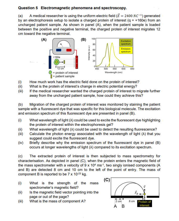
(a) A medical researcher is using the uniform electric field (E = 2400 NC-¹) generated
by an electrophoresis setup to isolate a charged protein of interest (q = +160e) from an
uncharged patient sample. As shown in panel (A), when the patient sample is loaded
between the positive and negative terminal, the charged protein of interest migrates 12
cm toward the negative terminal.
(A)
(B)
€€€
(ii)
(iii)
(iv)
(c)
+160e
-0
(1)
(ii)
(iii)
12 cm
= protein of interest
= patient sample
Intensity (a.u.)
100
80
60
40
20
ol
450
Excitation
spectrum
(b) Migration of the charged protein of interest was monitored by staining the patient
sample with a fluorescent dye that was specific for this biological molecule. The excitation
and emission spectrum of this fluorescent dye are presented in panel (B).
(1)
Emission
spectrum
500 550 600
Wavelength (nm)
How much work has the electric field done on the protein of interest?
What is the protein of interest's change in electric potential energy?
If the medical researcher wanted the charged protein of interest to migrate further
away from the uncharged patient sample, how could they achieve this?
What is the strength of the mass
spectrometer's magnetic field?
Is the magnetic field vector pointing into the
page or out of the page?
What is the mass of component A?
650
What wavelength of light (A) could be used to excite the fluorescent dye highlighting
the protein of interest within the electrophoresis gel?
What wavelength of light (A) could be used to detect the resulting fluorescence?
Calculate the photon energy associated with the wavelength of light (A) that you
suggest could excite the fluorescent dye.
Briefly describe why the emission spectrum of the fluorescent dye in panel (B)
occurs at longer wavelengths of light (A) compared to its excitation spectrum.
The extracted protein of interest is then subjected to mass spectrometry for
characterisation. As depicted in panel (C), when the protein enters the magnetic field of
the mass spectrometer with a velocity of 9 x 10⁴ ms-1, two singly ionised components (A
and B) are detected 8 cm and 10 cm to the left of the point of entry. The mass of
component B is reported to be 7 x 10-23 kg.
(C)
2 cm
A B
0024
8 cm Protein of
interest
Expert Solution
This question has been solved!
Explore an expertly crafted, step-by-step solution for a thorough understanding of key concepts.

Step by step
Solved in 3 steps with 7 images
