ACTIVITY 2 e your computer to view images that show cross-sections of compact bone. What you see should appear similar to Figure 6-2. A number of central canals should be visible in each image; these may appear either black or clear, depending on how the slide was prepared. Around cach central canal should be able to observe circular patterns of smaller black spots. Each of these spots is a lacuna, and in living bone an ostcocyte would be found within each lacuna. View some higher magnification images. You should be able to sce that each lacuna looks like a spider with many legs radiating from its body. The "legs" are the canaliculi, and you can sce how they connect the lacunae to cach other. Ironically, the dense, solid matrix appears clear when viewing it with a microscope. In the figure below, identify the following structures: central canal, lacuna, canaliculi, lamella. Use you Figure 6-2B. Compact bone: perforating canal. Figure 6-2A. Compact bone: osteon. OPGCC Deparment of Biological Sciences OPGCC Depament of Biological Scionces
ACTIVITY 2 e your computer to view images that show cross-sections of compact bone. What you see should appear similar to Figure 6-2. A number of central canals should be visible in each image; these may appear either black or clear, depending on how the slide was prepared. Around cach central canal should be able to observe circular patterns of smaller black spots. Each of these spots is a lacuna, and in living bone an ostcocyte would be found within each lacuna. View some higher magnification images. You should be able to sce that each lacuna looks like a spider with many legs radiating from its body. The "legs" are the canaliculi, and you can sce how they connect the lacunae to cach other. Ironically, the dense, solid matrix appears clear when viewing it with a microscope. In the figure below, identify the following structures: central canal, lacuna, canaliculi, lamella. Use you Figure 6-2B. Compact bone: perforating canal. Figure 6-2A. Compact bone: osteon. OPGCC Deparment of Biological Sciences OPGCC Depament of Biological Scionces

Anatomy & Physiology
1st Edition
ISBN:9781938168130
Author:Kelly A. Young, James A. Wise, Peter DeSaix, Dean H. Kruse, Brandon Poe, Eddie Johnson, Jody E. Johnson, Oksana Korol, J. Gordon Betts, Mark Womble
Publisher:Kelly A. Young, James A. Wise, Peter DeSaix, Dean H. Kruse, Brandon Poe, Eddie Johnson, Jody E. Johnson, Oksana Korol, J. Gordon Betts, Mark Womble
Chapter9: Joints
Section: Chapter Questions
Problem 1ILQ: Go to this website (http://openstaxcollege.org/l/childhand) to view a radiograph (X-ray image) of a...

Related questions
Question
100%
Can you help me with this activities and explain,
I can’t identify the structure.
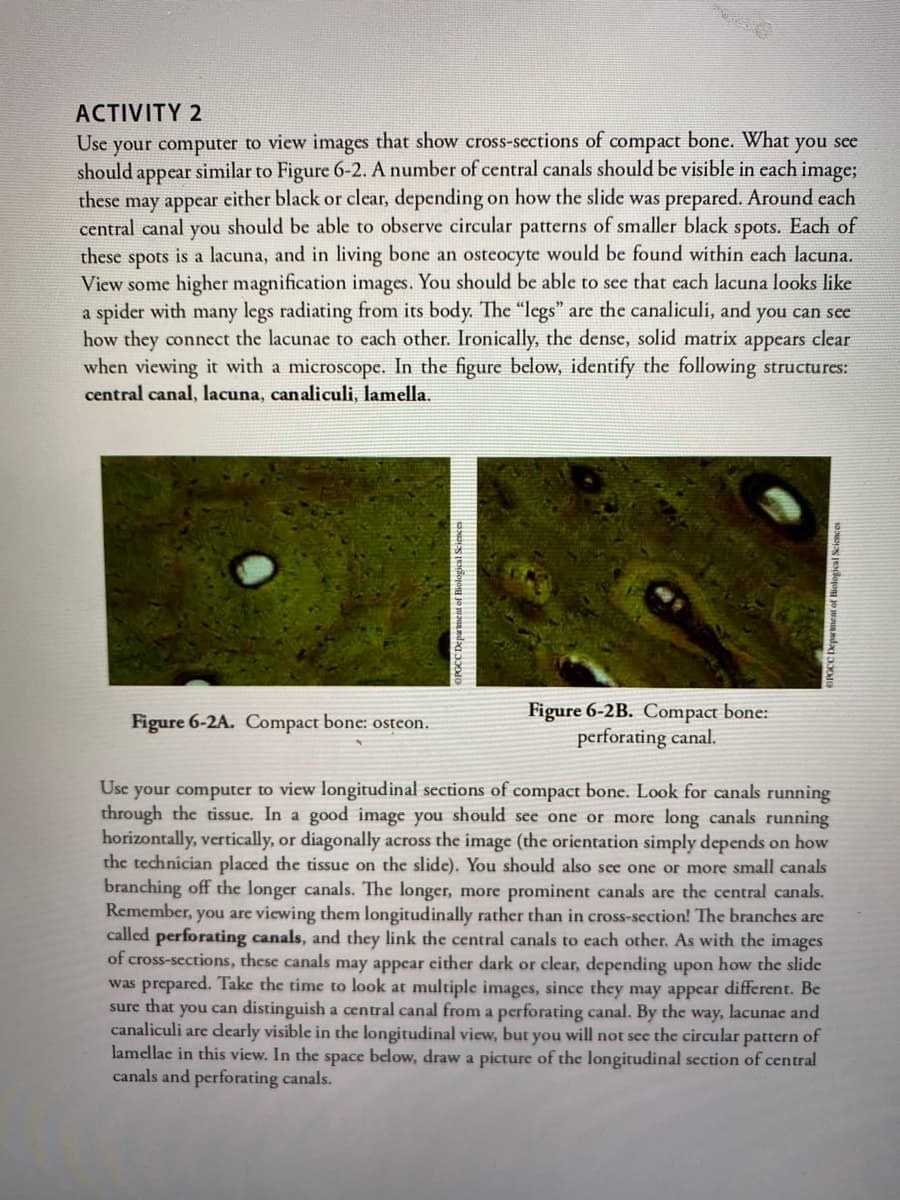
Transcribed Image Text:ACTIVITY 2
your computer to view images that show cross-sections of
should appear similar to Figure 6-2. A number of central canals should be visible in each image;
Use
compact
bone. What you see
these
either black or clear, depending
on how the slide was prepared. Around cach
may appear
central canal you should be able to observe circular patterns of smaller black spots. Each of
spots is a lacuna, and in living bone an osteocyte would be found within each lacuna.
View some higher magnification images. You should be able to sce that each lacuna looks like
a spider with many legs radiating from its body. The "legs" are the canaliculi, and you can sce
how they connect the lacunae to cach other. Ironically, the dense, solid matrix appears clear
when viewing it with a microscope. In the figure below, identify the following structures:
these
central canal, lacuna, canaliculi, lamella.
Figure 6-2B. Compact bone:
perforating canal.
Figure 6-2A. Compact bone: osteon.
Use your computer to view longitudinal sections of compact bone. Look for canals running
through the tissue. In a good image you should see one or more long canals running
horizontally, vertically, or diagonally across the image (the orientation simply depends on how
the technician placed the tissue on the slide). You should also see one or more small canals
branching off the longer canals. The longer, more prominent canals are the central canals.
Remember, you are viewing them longitudinally rather than in cross-section! The branches are
called perforating canals, and they link the central canals to cach other. As with the images
of cross-sections, these canals may appear either dark or clear, depending upon how the slide
was prepared. Take the time to look at multiple images, since they may appear different. Be
sure that you can distinguish a central canal from a perforating canal. By the way, lacunae and
canaliculi are dearly visible in the longitudinal view, but you will not see the circular pattern of
lamellae in this view. In the space below, draw a picture of the longitudinal section of central
canals and perforating canals.
OPGCC Department of Biological Scicnces
Biological Sciences
Expert Solution
This question has been solved!
Explore an expertly crafted, step-by-step solution for a thorough understanding of key concepts.

This is a popular solution!

Trending now
This is a popular solution!

Step by step
Solved in 3 steps with 1 images

Recommended textbooks for you
Anatomy & Physiology
Biology
ISBN:
9781938168130
Author:
Kelly A. Young, James A. Wise, Peter DeSaix, Dean H. Kruse, Brandon Poe, Eddie Johnson, Jody E. Johnson, Oksana Korol, J. Gordon Betts, Mark Womble
Publisher:
OpenStax College

Biology 2e
Biology
ISBN:
9781947172517
Author:
Matthew Douglas, Jung Choi, Mary Ann Clark
Publisher:
OpenStax

Anatomy & Physiology
Biology
ISBN:
9781938168130
Author:
Kelly A. Young, James A. Wise, Peter DeSaix, Dean H. Kruse, Brandon Poe, Eddie Johnson, Jody E. Johnson, Oksana Korol, J. Gordon Betts, Mark Womble
Publisher:
OpenStax College
Biology 2e
Biology
ISBN:
9781947172517
Author:
Matthew Douglas, Jung Choi, Mary Ann Clark
Publisher:
OpenStax

Medical Terminology for Health Professions, Spira…
Health & Nutrition
ISBN:
9781305634350
Author:
Ann Ehrlich, Carol L. Schroeder, Laura Ehrlich, Katrina A. Schroeder
Publisher:
Cengage Learning

Human Biology (MindTap Course List)
Biology
ISBN:
9781305112100
Author:
Cecie Starr, Beverly McMillan
Publisher:
Cengage Learning