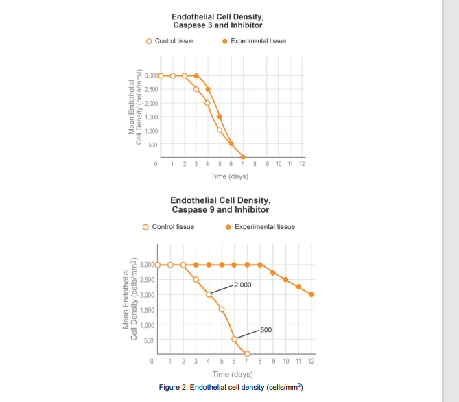
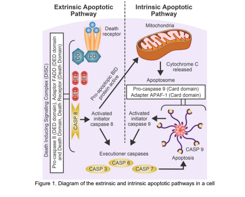
Caspase proteins are enzymes known to play a role in programmed cell death (apoptosis) and  the inflammatory response. Apoptotic caspases are subcategorized into initiator and executioner  caspases. Initiator caspases produce a chain reaction that activates executioner caspases.  Caspase 9 is a kind of initiator caspase and caspase 3 is a kind of executioner caspase that plays  a direct role in degrading cellular components. Apoptosis can be activated by internal (intrinsic) cellular mechanisms or external (extrinsic)  signals. The extrinsic apoptotic pathway begins with the reception of a signal at the death  receptors and the intrinsic apoptotic pathway begins with the permeabilization of the  mitochondria. Both apoptotic caspase pathways are shown in Figure 1. Caspase proteins have been implicated in the premature death of cornea endothelial tissue being  stored for transplant. To investigate the effect of caspases 3 and 9 on tissue degradation,  scientists monitored the endothelial cell density of cornea tissue, left isolated from other tissues,  over a period of seven days. Scientists then exposed cornea endothelial tissue to caspase 3 and  caspase 9 inhibitors and monitored the endothelial tissue cell density over a period of 12 days  (Figure 2).
Anatomy & Physiology
1st Edition
ISBN:9781938168130
Author:Kelly A. Young, James A. Wise, Peter DeSaix, Dean H. Kruse, Brandon Poe, Eddie Johnson, Jody E. Johnson, Oksana Korol, J. Gordon Betts, Mark Womble
Publisher:Kelly A. Young, James A. Wise, Peter DeSaix, Dean H. Kruse, Brandon Poe, Eddie Johnson, Jody E. Johnson, Oksana Korol, J. Gordon Betts, Mark Womble
Chapter10: Muscle Tissue
Section: Chapter Questions
Problem 23RQ: Which of the following statements is false? Slow fibers have a small network of capillar ies. Slow...
icon
Related questions
Question

Caspase proteins are enzymes known to play a role in programmed cell death (apoptosis) and 
the inflammatory response. Apoptotic caspases are subcategorized into initiator and executioner 
caspases. Initiator caspases produce a chain reaction that activates executioner caspases. 
Caspase 9 is a kind of initiator caspase and caspase 3 is a kind of executioner caspase that plays 
a direct role in degrading cellular components.
Apoptosis can be activated by internal (intrinsic) cellular mechanisms or external (extrinsic) 
signals. The extrinsic apoptotic pathway begins with the reception of a signal at the death 
receptors and the intrinsic apoptotic pathway begins with the permeabilization of the 
mitochondria. Both apoptotic caspase pathways are shown in Figure 1.

Caspase proteins have been implicated in the premature death of cornea endothelial tissue being 
stored for transplant. To investigate the effect of caspases 3 and 9 on tissue degradation, 
scientists monitored the endothelial cell density of cornea tissue, left isolated from other tissues, 
over a period of seven days. Scientists then exposed cornea endothelial tissue to caspase 3 and 
caspase 9 inhibitors and monitored the endothelial tissue cell density over a period of 12 days 
(Figure 2).
 

Endothelial Cell Density,
Caspase 3 and Inhibitor
Control tissue
Experimental tissue
3,0000
2,500
2,000
1,500
1,000
500
1 2 3 4 5 678 9 10 11 12
Time (days)
Endothelial Cell Density,
Caspase 9 and Inhibitor
O Control tissue
Experimental tissue
3,0000
2,500
-2,000
2,000
1,500
1,000
-500
500
0 1 2 3 4 5 6 7 8 9 10 11 12
Time (days)
Figure 2. Endothelial cell density (cells/mm?)
Mean Endothelial
Mean Endothelial
Cell Density (cells/mm2)
Transcribed Image Text:Endothelial Cell Density, Caspase 3 and Inhibitor Control tissue Experimental tissue 3,0000 2,500 2,000 1,500 1,000 500 1 2 3 4 5 678 9 10 11 12 Time (days) Endothelial Cell Density, Caspase 9 and Inhibitor O Control tissue Experimental tissue 3,0000 2,500 -2,000 2,000 1,500 1,000 -500 500 0 1 2 3 4 5 6 7 8 9 10 11 12 Time (days) Figure 2. Endothelial cell density (cells/mm?) Mean Endothelial Mean Endothelial Cell Density (cells/mm2) Cell Density (cells/mm2)
Extrinsic Apoptotic
Pathway
Intrinsic Apoptotic
Pathway
Mitochondria
Death
receptor !
DD
Cytochrome C
released
Apoptosome
Pro-caspase 9 (Card domain)
Adapter APAF-1 (Card domain)
i Activated
initiator
! caspase 9
Activated
initiator
caspase 8
CASP 9
Executioner caspases
Арoptosis
CASP 6
CASP 3
CASP 7
Figure 1. Diagram of the extrinsic and intrinsic apoptotic pathways in a cel
cll
Pro-caspase 8 (DED domain), Adaptor FADD (DED domain
and Death Domain, Death Receptor (Death Domain)
DED
Death Inducing Signaling Complex (DISC)
DED
DED
aa
CASP 8
Pro-apoptopic BID
protein active
Transcribed Image Text:Extrinsic Apoptotic Pathway Intrinsic Apoptotic Pathway Mitochondria Death receptor ! DD Cytochrome C released Apoptosome Pro-caspase 9 (Card domain) Adapter APAF-1 (Card domain) i Activated initiator ! caspase 9 Activated initiator caspase 8 CASP 9 Executioner caspases Арoptosis CASP 6 CASP 3 CASP 7 Figure 1. Diagram of the extrinsic and intrinsic apoptotic pathways in a cel cll Pro-caspase 8 (DED domain), Adaptor FADD (DED domain and Death Domain, Death Receptor (Death Domain) DED Death Inducing Signaling Complex (DISC) DED DED aa CASP 8 Pro-apoptopic BID protein active
Expert Solution
trending now

Trending now

This is a popular solution!

steps

Step by step

Solved in 2 steps

Blurred answer
Knowledge Booster
Genomic studies
Learn more about
Need a deep-dive on the concept behind this application? Look no further. Learn more about this topic, biology and related others by exploring similar questions and additional content below.
Similar questions
  • SEE MORE QUESTIONS
Recommended textbooks for you
Anatomy & Physiology
Biology
ISBN:
9781938168130
Author:
Kelly A. Young, James A. Wise, Peter DeSaix, Dean H. Kruse, Brandon Poe, Eddie Johnson, Jody E. Johnson, Oksana Korol, J. Gordon Betts, Mark Womble
Publisher:
OpenStax College