Draw a plan diagram of the frog intestine viewed under the microscope (see attached images for help!!).... A plan diagram is a simple drawing showing only the boundaries between the lumen, the columnar epithelium, the (areolar) connective tissue, the circular smooth muscle and the longitudinal smooth muscle. A plan diagram serves to illustrate the location and relative thickness of the various tissue layers. 1. Draw a plan diagram and label the following: • Longitudinal smooth muscle • Columnar epithelial tissue • Circular smooth muscle • Villi • Areolar connective tissue • Lumen 2. Calculate actual size and drawing magnification of the width of the intestine. Include formulae used and calculations. 3. Add a scale bar with actual size next to your diagram. Give your drawing a descriptive title and record total magnification. Attached are an example plan diagram labelled, as well as a microscopic image of the frog intestine (3-4 cells should fit across only) PLEASE DO THE DRAWING AND ANSWER QUESTION CORRECTLY ?


Draw a plan diagram of the frog intestine viewed under the microscope (see attached images for help!!)....
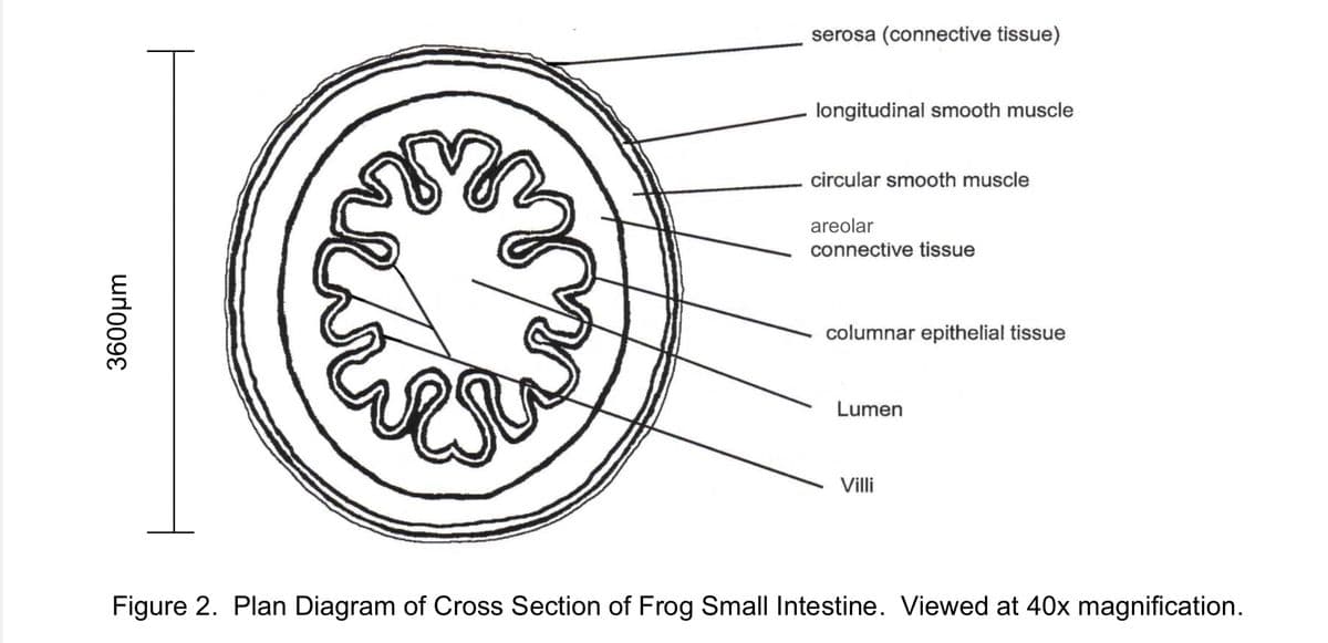
A plan diagram is a simple drawing showing only the boundaries between the lumen, the columnar epithelium, the (areolar) connective tissue, the circular smooth muscle and the longitudinal smooth muscle. A plan diagram serves to illustrate the location and relative thickness of the various tissue layers.
1. Draw a plan diagram and label the following: • Longitudinal smooth muscle • Columnar epithelial tissue • Circular smooth muscle • Villi • Areolar connective tissue • Lumen
2. Calculate actual size and drawing magnification of the width of the intestine. Include formulae used and calculations.
3. Add a scale bar with actual size next to your diagram.
Give your drawing a descriptive title and record total magnification.
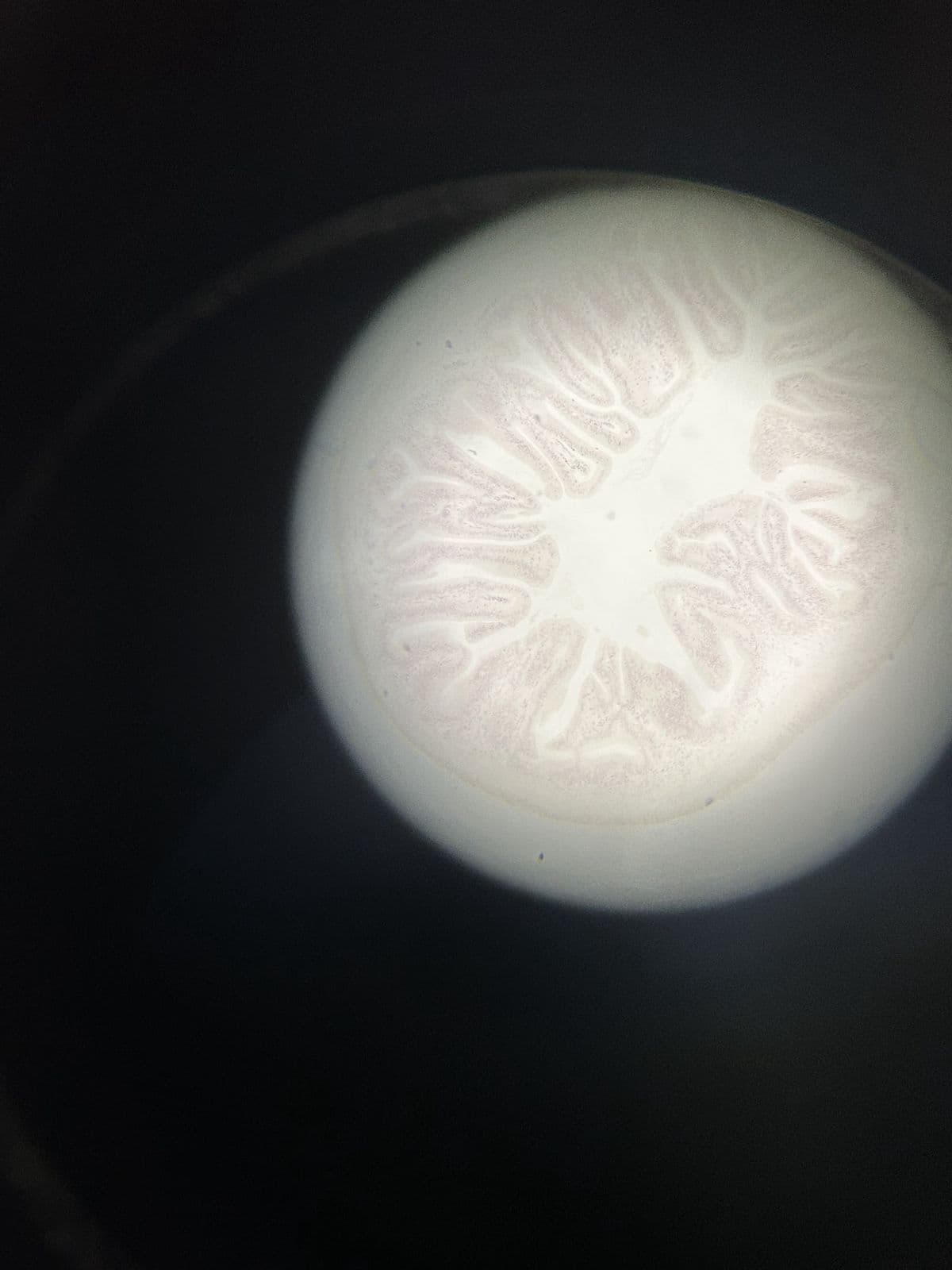
Attached are an example plan diagram labelled, as well as a microscopic image of the frog intestine (3-4 cells should fit across only)
PLEASE DO THE DRAWING AND ANSWER QUESTION CORRECTLY ?


Trending now
This is a popular solution!

Step by step
Solved in 3 steps with 1 images


