

Q: D E- F- Zygomatic Arch Dental Arcade Foramen Magnum Occipital Condyle Occipital Bone [Choose ]…
A: The skeleton of the head is called the skull. It consists of several bones that are joined together…
Q: al ULNA CERVICLE RIB SKULL
A: Bone consists of hard connective tissue called osseous. Functions of bone Protecting vital organs…
Q: Chosse correct option
A: As infants grow, their food requirements also change as per the need of the body. The infants should…
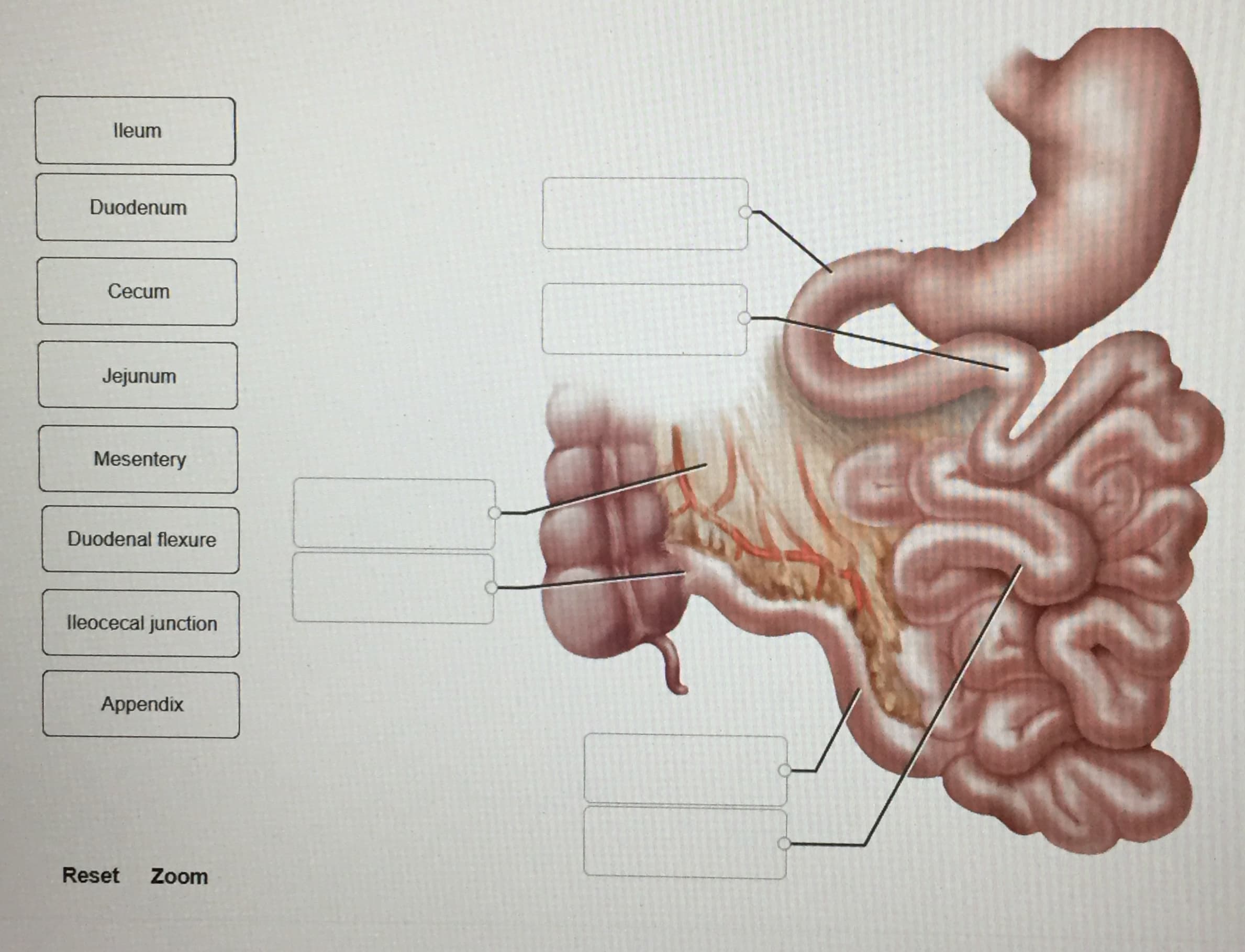
Q: Possible function for appendix, muscles for moving ear, body hair, little toe, and tailbone?
A: Each organ has some function to assist with even if not a major contribution to the system overall.
Q: Difference of contusions from concussions
A: One might assume a concussion is just a more serious contusion. But the reality is that both…
Q: Select one: Oa soft palate Ob. sphenoidal sinus Oc external nares
A: Anatomy is a discipline of biology concerned with the characterization of bodily structures revealed…
Q: femoral nerve inferior gluteal nerve superior gluteal nerve tibial nerve
A: Answering only first question. Kindly repost second question to be answered Knee stiffness can…
Q: Type of muscle: Structure B:
A: Ans. The figure represents the structure of the cardiac muscle. Muscle is an organ made up of muscle…
Q: Terms Sternocleidomastoid Characteristics Biceps Attachment Sites Fiber Direction Number of Heads…
A: Muscle is a type of contractile tissue that is organized into coordinated systems for increased…
Q: ORIGIN OF CRANIOFACIAL MUSCLE SOMITΕ ΝΟ. ΑΝD MUSCLE INNEVAΤION MESODERMAL ORIGIN 1 2 3 4 7
A: Introduction: Craniofacial muscles or facial muscles comprising of 20flat skeletal muscles lying…
Q: Label the bone markings listed in Figure
A: The figure given is the floor of the cranial cavity. The cranial cavity is the space within the…
Q: Label the: Proximal extensor retinaculum Long lateral collateral ligament Short lateral collateral…
A: Muscles, tendons, and ligaments run over the surfaces of the foot, allowing for sophisticated…
Q: C Caudal Crania
A: Anatomy A field of biology which deals with the structure of the organ and their part of a…
Q: Structure Location Function Nicticiating Membrane Vomerine Teeth Maxillary Teeth Eustacian Tubes…
A: The location and function of different parts of frog are as follows:
Q: Extensors Flexors Levators Depressors Compressors Quadratus lumborum Semispinalis cervicis Longus…
A: Anatomy and physiology are the branch of biology, anatomy deals with the study of the structure of…
Q: Superficial Beep Gluteus maximus Latissimus dorsi Thoracolumbar fascia Deltoid Sternocleiodmastoid
A: Gluteus maximus : It is the major extensor of the hip, out of the three gluteal muscle this one is…
Q: Movement of the thumb across the hand to meet the little finger is Radial deviation
A: The developments at each thumb joint are flexion and expansion and extra developments of…
Q: he femoral canal?
A: Femoral canal : It is an anatomical compartment located in the anterior thigh . It is the smallest…
Q: Suprascapular carțilage Acetabular surface Puboischiadic bar Proptergium Scapular cartilage…
A: The suppprtimg skeleton of the fins constitutes the appendicular skeleton. Two appendicular skeleton…
Q: What is the subcortical stucture and function
A: Subcortical structures are a type of diverse neural formations deep within the brain which include…
Q: Musculo skeletal system Physical Examination: Shoulders: Active range of motion: Adduction:…
A: Physical examination- Inspection and palpation are the two main primary methods used for physical…
Q: What is the difference between the Pars Recta and the Pars Oblique?
A: The cricothyroid cartilage has a fan-shaped structure and it diverges into - a)Par recta and b) Pars…
Q: mylohyoid 11 12 13 pectoralis sternalis transversus linea alba 14 Gur
A: The given image shows the musculature of the ventral side of a frog. It is required to identify the…
Q: Bob is running hurdles and suffers an injury that causes his hamstring to roll up into his gluteus…
A: The gluteus muscles are of three types, namely, gluteus medius, gluteus maximus, and gluteus…
Q: Define flatus
A: Different body organs work, in a coordinated way to maintain optimum body functioning.…
Q: ·松 Name Section Date 9 Label the following parts of the shoulder joint in Figure 9.10. ■ Biceps…
A: Image A –Joint capsule – It is also termed as articular capsule. It surrounds synovial joint.Glenoid…
Q: anterior drawer sign. An MRI shows injury to several structures of the knee, including her medial…
A: Injuries occurring due to lateral blue to the knees can be tears in the lateral collateral ligament.…
Q: Fibrocartilage (____×)Location _______________________ Function _______________________
A: As we know human body is made of skeleton. Skeleton is structure that keeps body in shape and helps…
Q: Label the Figure
A: The blood is circulated throughout the body parts by the circulatory system whose main parts include…
Q: to prep abdominal area for surgery.
A: Preoperative preparation Proper preoperative preparation optimizes postoperative outcomes. Routine…
Q: Medical intervention of post abdomenal weakness
A: The key to recovering after a caesarean section or an open hernia repair is to move slowly. After…
Q: O fascicle
A: Muscles and nerves are an important part of the body required for locomotion, movement as well as…
Q: The calcaneal region is _____ to the cervical region.
A: The anatomical position is a reference point which is used to mention the location of the body…
Q: What option does the patient have to replace the missing teeth in the mandibular arch
A: Dentistry It deals with dental medicine and oral medicine. It consists of the study, diagnosis,…
Q: G0:6 a docs.google.com Examine the figure carefully and then name the labeled numbers: 12 10 11 3. 4…
A: Both the tendons and ligaments provide stability to the bones and play an important role in the…
Q: Axial Skeleton Skull Facial Bones Maxillae Anterior Nasal Spine Alveolar Processes Infraorbital…
A: Answer
Q: Activity D- Label the regions of the abdomen 4. Label the 4 quadrants of the abdominal area
A: 4. The four quadrants of the abdominal area are- Right upper Quadrant Left upper Quadrant…
Q: Can you help me the name of the muscles of the labels?
A: In this question, we have to name the muscles of labels. I labelled all visible labelling of the…
Q: Postanal tail Cranium
A: Answer Chordata has four characteristics that differ from non chordata 1. Dorsal hollow nerve cord…
Q: cervical spine and upper limb major musclesss
A: Cervical bones consists of 7 vertebral bodies C1 - C7 and are separated by intervertebral disc.…
Q: (Aca) Anterior champer angle p 2:36
A: Anterior champer angle (ACA) is the healthy angle of eye and it is located between the Iris and…
Q: Copyright Nielsen & Miller O accessory (XI) nerve vestibulochoclear (VIII) nerve O Hypoglossal (XII)…
A: The correct answer to this particular question will be, accessory (XI) nerve
Q: temporalis 2 7 A. mentalis B. sternocleidoastoid C. buccinator D. platysma E. masseter F.…
A:
Q: Figure: Lateral view of the right shoulder and upper limb. A. Bone B. Landmark B C. Landmark D.…
A: A joint is an articulation between two bones in the body and is comprehensively classified by the…
Q: Frontal Sinus 45 46 Ethmoid bone nternal Acoustic meatus Internal Auditory Meatus 47 48 Sphenoid…
A: The above diagram shows the lateral view of skull with certain prominent landmarks and the second…
Q: Gastrocnemius Fibularis longus Soleus Tibialis anterior Extensor digitorum longus
A: Gartrocnemius muscle is a two headed Muscle which is present in the back portion of lower leg .…
Q: usculo skeletal examination: Cervical Spine 1. Contour: 2. Mass or deformity: 3. Pain: 4.…
A: Components included for the physical examination are as follows:-Inspection -Palpation -Percussion…


Trending now
This is a popular solution!

Step by step
Solved in 3 steps with 1 images
