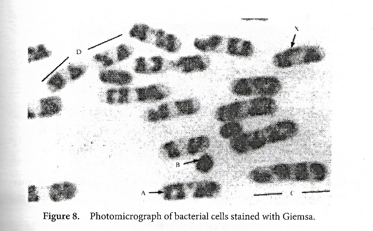
Measure the length of Cell X using the ruler in Microsoft Word. Assuming the actual length to be 3 um, calculate the magnification. Show your complete solution. Based on Figure 1, what internal organization can be distinguished in cell X? Why? Based in Figure 1, can you see a limiting membrane? Can you deduce its presence? Why or why not? Figure 2 is an electron micrograph of the same type of bacterium as shown in Figure 1. The picture has been obtained by cutting a very thin section of the bacterial cell along its longitudinal axis. Measure the total length of the cell, and assuming the actual length to be 2.1 um, calculate the magnification. With reference with Figure 2, what are the major differences between the inclusions found in Figure 1 and the way they appear in Figure 2? What other structural features can be resolved?


Structure and Composition of Cell Membrane
Despite differences in structure and function, all living cells in multicellular organisms are surrounded by a cell membrane. Just like the outer layer of the skin separates the body from its environment similarly, the cell membrane, also known as 'plasma membrane,' separates the inner content from its exterior environment.
Cell Membrane
The cell membrane is known by different names like plasma membrane or cytoplasmic membrane, or biological membrane. The term "cell membrane" was first introduced by C. Nageli and C. Cramer in the year 1855. Later on, in 1931, the term "plasmalemma" for cell membrane was given by J. Plowe. The cell membrane separates the cell's internal environment from the extracellular space. This separation allows the protection of cells from their environment.
Prokaryotes vs Eukaryotes
The cell is defined as the basic structural and functional unit of life. The cell membrane bounds it. It is capable of independent existence.
Measure the length of Cell X using the ruler in Microsoft Word. Assuming the actual length to be 3 um, calculate the magnification. Show your complete solution.
- Based on Figure 1, what internal organization can be distinguished in cell X? Why?
- Based in Figure 1, can you see a limiting membrane? Can you deduce its presence? Why or why not?
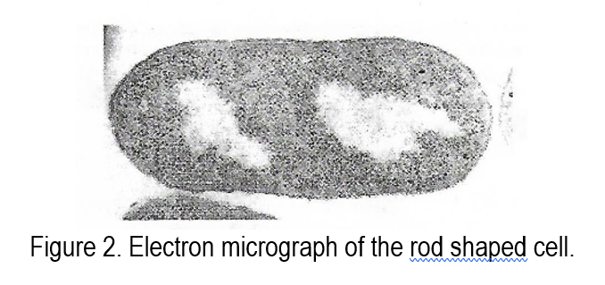
Figure 2 is an electron micrograph of the same type of bacterium as shown in Figure 1. The picture has been obtained by cutting a very thin section of the bacterial cell along its longitudinal axis.
Measure the total length of the cell, and assuming the actual length to be 2.1 um, calculate the magnification.
- With reference with Figure 2, what are the major differences between the inclusions found in Figure 1 and the way they appear in Figure 2?
- What other structural features can be resolved?

Step by step
Solved in 3 steps


