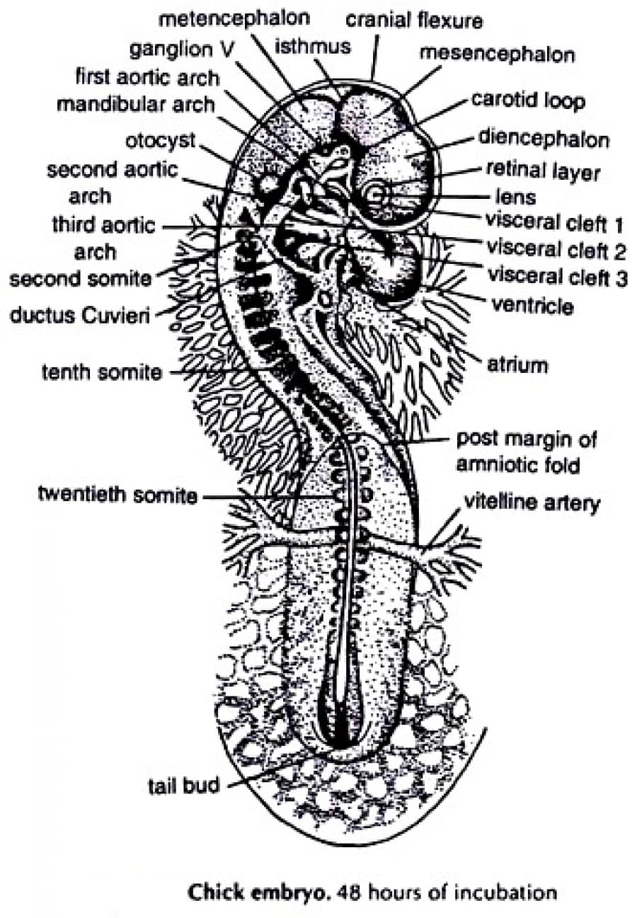
metencephalon isthmus cranial flexure ganglion V first aortic arch mandibular arch mesencephalon carotid loop otocyst. second aortic arch diencephalon retinal layer -lens visceral cleft 1 visceral cleft 2 visceral cleft 3 third aortic arch second somite ventricle ductus Cuvieri atrium tenth somite - post margin of amniotic fold twentieth somite vitelline artery


The 48-hr Chick Embryo Whole Mount (SEE IMAGE)
Before you mount your slide on your microscope stage, take a good look at the morphology of the 48-hour embryo. Note the striking changes in morphology as compare, to the 33-hour embryo. There is a cephalic flexure or bending at the level of the midbrain. The head now lies on its left side. On the other hand dorsal side of the trunk is still turned up so the twisting of the embryo is noticeable.
Verify these changes under the low power objective. Note the flexion and the torsion. In addition to the cranial/cephalic flexure there is also a cervical flexure at the level of the neck region.
Very briefly go through the structures previously seen in the 33-hour chick. Observe the new structures appearing for the first time (Please visit the main website for viewing the whole mount of 33-hour and 48-hour chick embryo).
Ectodermal Derivatives
The prosencephalon is now divided into two regions:
Telencephalon: This brain region is the anterior division of the prosencephalon. Look for the landmarks: the velum transversum, a depression in the roof of the forebrain and the optic recess, a slight ventral notch marking the demarcation between the telencephalon and the diencephalon.
Diencephalon: The posterior division of the prosencephalon is the diencephalon. This is distinguished by the optic cup which are its lateral expansions.
Mesencephalon: The midbrain is the site of the cephalic flexure. The isthmus is the constriction between the mesencephalon and the metencephalon.
Meso-diencephalic fold: This is the dorsal constriction between the diencephalon and the mesencephalon.
Tuberculum posterius: This ventral elevation is opposite the meso- diencephalic fold.
Metencephalon: The brain region posterior to the mesencephalon is the metencephalon. Anteriorly it is delimited by the isthmus and posteriorly by the thinning out of the roof, which is characteristic of the next brain region.
Myelencephalon: This is the brain region posterior to the metencephalon. The thin roof sinks into the myelocoel to become the posterior choroid plexus.
Eyes: Identify the optic cup with its two layers, and the lens vesicle.
Inner ear: What appears as a dorsal evagination of the otocyst is the forerunner of the endolymphatic duet.
Endodermal Derivatives
Digestive Tract
At this stage there is now a pharynx with the characteristic pharyngeal arches found ventral to the myelencephalon. The first or hyomandibular arch is bounded posteriorly by the hyomandibular groove. The second visceral or hyoid arch is bounded in front by this groove and behind by the second groove. Can you see a third visceral arch? Observe the aortic arches, the tiny clear vessels running through each arch.
Anterior intestinal portal: This is the opening of the foregut into the yolk
Posterior intestinal portal: This is the opening of the hindgut into the yolk
Hindgut: This sacculation of the endoderm into the tail bud is the hindgut.
What other digestive tract derivatives can you see in whole mount?
Mesodermal Derivatives
Circulatory System
Heart: This organ has twisted upon itself due to its elongation in a limited space. The ventricle now lies posterior to the atrium and both lie outside the body of the embryo. Quickly identify the structures that you had earlier seen in the 33- hour chick embryo
Common cardinal veins or ducts of Cuvier: These paired short vessels receive the anterior cardinal and posterior cardinal veins before they enter the sinus venosus.
Somites: Count the number of somites at this stage. Identify the regions of the somite, the dermo-myotome plate and the sclerotome.
Tail bud: This cell mass is at the posterior tip of the embryo. It marks the position of the Hensen's node and the old primitive streak
Amniotic fold: This hood grows caudally over the embryo. It is composed of two membranes, the outer chorion and the inner amnion.
QUESTION:
- How are the °lacysts or auditory vesicles formed?
- At this stage of development, what cranial ganglia are present?


Trending now
This is a popular solution!

Step by step
Solved in 3 steps


