OBSERVATIONS AND INTERPRETATIONS 1 Record your observations in the table below. temitayo Cellular Morphology and Arrangement (include a written description and a detailed sketch of a few representative cells) Stain Organism and Duration Cell Dimensions
OBSERVATIONS AND INTERPRETATIONS 1 Record your observations in the table below. temitayo Cellular Morphology and Arrangement (include a written description and a detailed sketch of a few representative cells) Stain Organism and Duration Cell Dimensions

Essentials of Pharmacology for Health Professions
7th Edition
ISBN:9781305441620
Author:WOODROW
Publisher:WOODROW
Chapter6: Safe Dosage Calculations
Section: Chapter Questions
Problem B.4CRQ

Related questions

Question
Transcribed Image Text:OBSERVATIONS AND INTERPRETATIONS
1 Record your observations in the table below.
temitayo Cellular Morphology and Arrangement
(include a written description and a detailed sketch
of a few representative cells)
Stain
Organism
and Duration
Cell Dimensions
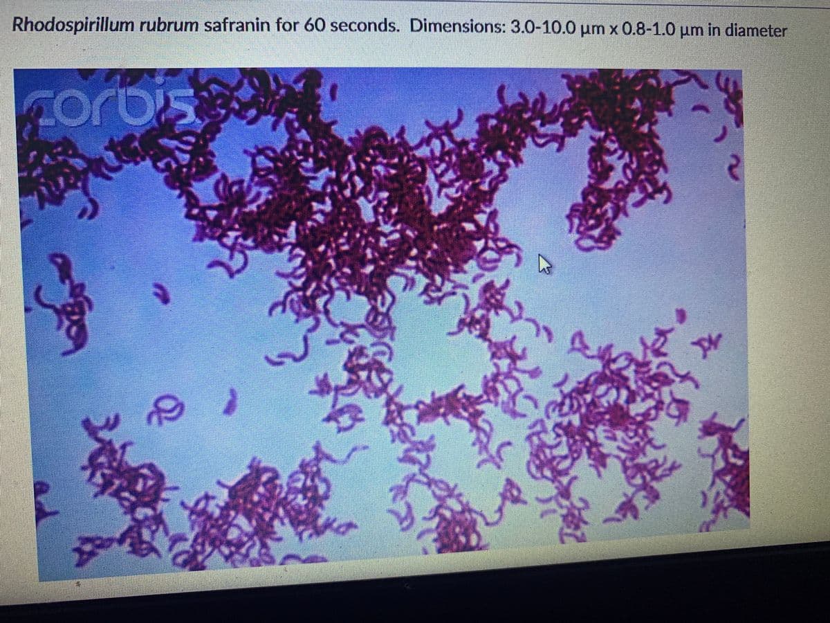
Transcribed Image Text:Rhodospirillum rubrum safranin for 60 seconds. Dimensions: 3.0-10.0 um x 0.8-1.0 µm in diameter
475
corbis
Expert Solution
Step 1
In a simple staining procedure, only one stain is used to stain the organism which produces a contrast when observed against an unstained background. The stains chosen for the simple staining technique are basic stains that positively charged chromogen so that it binds to the negatively charged nucleic acids and certain cell wall components of the organism. Examples of simple stains include methylene blue, crystal violet, and safranin. The morphology of the organism and the way in which they are arranged can be observed by a simple staining technique.
The procedure for simple staining technique is as follows:
- Clean the microscopic slides and dry them properly.
- Sterilize the inoculating loop and transfer a loopful of tap water on to the slide.
- Sterilize the inoculating loop and transfer a loopful of the inoculum onto the slide.
- Make a smear of the inoculum in the tap water.
- Heat fix the smear by passing the underside of the slide through the flame 3 to 4 times.
- Stain the slide with safranin and allow it to stay for 60 seconds.
- Wash off the excess stain and dry the slide
- Observe the slide under the microscope

Trending now
This is a popular solution!

Step by step
Solved in 2 steps with 1 images

Knowledge Booster


Learn more about
Need a deep-dive on the concept behind this application? Look no further. Learn more about this topic, biology and related others by exploring similar questions and additional content below.Recommended textbooks for you
Essentials of Pharmacology for Health Professions
Nursing
ISBN:
9781305441620
Author:
WOODROW
Publisher:
Cengage
Essentials of Pharmacology for Health Professions
Nursing
ISBN:
9781305441620
Author:
WOODROW
Publisher:
Cengage