Comment Figure 2. To facilitate your analysis, focus your comments on the squared region, where cells are the most representative

Human Biology (MindTap Course List)
11th Edition
ISBN:9781305112100
Author:Cecie Starr, Beverly McMillan
Publisher:Cecie Starr, Beverly McMillan
Chapter16: Reproductive Systems
Section: Chapter Questions
Problem 1SQ: Besides producing gametes (sperm and eggs), the primary male and female reproductive organs also...
icon
Related questions
Question

The single uterus (womb) anchors and nourishes the developing fetus. Its walls are composed of 3 layers of tissues, respectively the perimetrium (outer thin cover), the myometrium (thick layer 2 of smooth muscles, which contract to deliver the baby), and the endometrium (inner glandular
tissue that lines the internal uterus cavity and undergoes cyclical changes during the menstrual cycle). The uterus lower part is called the cervix and its cavity is called the cervical canal.
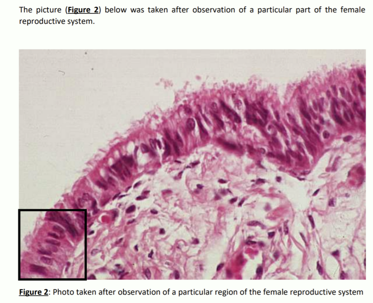
The oviducts, also called the Fallopian tubes (10-12 cm long), transport the ovocytes released from the ovaries to the uterus by peristalsis. The oviducts are not attached to the ovaries. Instead, the exhibit a funnel-shaped extremity, dubbed the infundibulum, that opens with finger-like
projections to the ovaries in order to collect the emitted ovocyte. The remaining parts of the Fallopian tubes are named respectively the ampulla (the longest part of a Fallopian tube), and the isthamus(the part of the Fallopian tube, which is closer to the uterus). Each Fallopian tube is lined
with cilia that help the ovocytes move to the uterus. If sperms are present, fertilization takes place in the upper end of the fallopian tubes, i.e. the ampulla.

Comment Figure 2. To facilitate your analysis, focus your comments on the squared
region, where cells are the most representative

The picture (Figure 2) below was taken after observation of a particular part of the female
reproductive system.
Figure 2: Photo taken after observation of a particular region of the female reproductive system
Transcribed Image Text:The picture (Figure 2) below was taken after observation of a particular part of the female reproductive system. Figure 2: Photo taken after observation of a particular region of the female reproductive system
Expert Solution
steps

Step by step

Solved in 2 steps

Blurred answer
Knowledge Booster
Developmental biology
Learn more about
Need a deep-dive on the concept behind this application? Look no further. Learn more about this topic, biology and related others by exploring similar questions and additional content below.
Similar questions
  • SEE MORE QUESTIONS
Recommended textbooks for you
Human Biology (MindTap Course List)
Biology
ISBN:
9781305112100
Author:
Cecie Starr, Beverly McMillan
Publisher:
Cengage Learning
Biology 2e
Biology 2e
Biology
ISBN:
9781947172517
Author:
Matthew Douglas, Jung Choi, Mary Ann Clark
Publisher:
OpenStax
Concepts of Biology
Concepts of Biology
Biology
ISBN:
9781938168116
Author:
Samantha Fowler, Rebecca Roush, James Wise
Publisher:
OpenStax College
Human Physiology: From Cells to Systems (MindTap …
Biology
ISBN:
9781285866932
Author:
Lauralee Sherwood
Publisher:
Cengage Learning