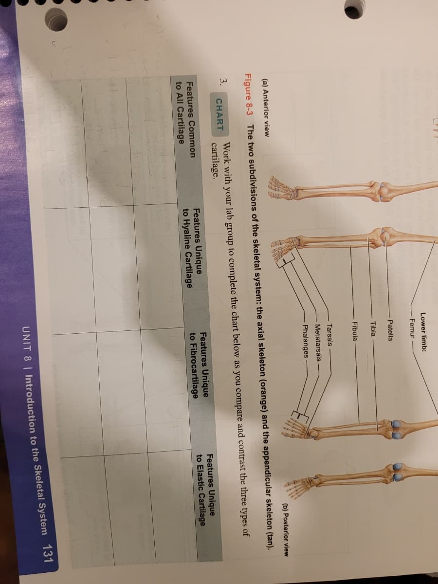
Compare and contrast the three types of cartilage

Anatomy & Physiology
1st Edition
ISBN:9781938168130
Author:Kelly A. Young, James A. Wise, Peter DeSaix, Dean H. Kruse, Brandon Poe, Eddie Johnson, Jody E. Johnson, Oksana Korol, J. Gordon Betts, Mark Womble
Publisher:Kelly A. Young, James A. Wise, Peter DeSaix, Dean H. Kruse, Brandon Poe, Eddie Johnson, Jody E. Johnson, Oksana Korol, J. Gordon Betts, Mark Womble
Chapter7: Axial Skeleton
Section: Chapter Questions
Problem 18RQ: A typical vertebra has ________. a vertebral foramen that passes through the body a superior...
icon
Related questions
icon
Concept explainers
Question
100%
Compare and contrast the three types of cartilage
Lower limb:
Femur
Patella
Tibia
Fibula
Tarsals
Metatarsals
Phalanges
(b) Posterior view
(a) Anterior view
Figure 8-3 The two subdivisions of the skeletal system: the axial skeleton (orange) and the appendicular skeleton (tan).
Work with your lab group to complete the chart below as you compare and contrast the three types of
cartilage.
3.
CHART
Features Unique
to Hyaline Cartilage
Features Unique
to Fibrocartilage
Features Unique
to Elastic Cartilage
Features Common
to All Cartilage
UNIT 8 | Introduction to the Skeletal System 131
Transcribed Image Text:Lower limb: Femur Patella Tibia Fibula Tarsals Metatarsals Phalanges (b) Posterior view (a) Anterior view Figure 8-3 The two subdivisions of the skeletal system: the axial skeleton (orange) and the appendicular skeleton (tan). Work with your lab group to complete the chart below as you compare and contrast the three types of cartilage. 3. CHART Features Unique to Hyaline Cartilage Features Unique to Fibrocartilage Features Unique to Elastic Cartilage Features Common to All Cartilage UNIT 8 | Introduction to the Skeletal System 131
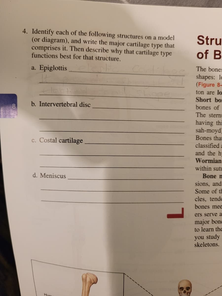
4. Identify each of the following structures on a model
(or diagram), and write the major cartilage type that
comprises it. Then describe why that cartilage type
functions best for that structure.
Stru
of B
a. Epiglottis
The bones
shapes: lo
(Figure 8-
ton are lo
Short bor
b. Intervertebral disc
bones of
The stern
having thi
sah-moyd)
Bones that
classified a
c. Costal cartilage
and the h
Wormian
within sutu
d. Meniscus
Bone m
sions, and
Some of th
cles, tendo
bones mee
ers serve a.
major bone
to learn the
you study
skeletons.
Hum
Transcribed Image Text:4. Identify each of the following structures on a model (or diagram), and write the major cartilage type that comprises it. Then describe why that cartilage type functions best for that structure. Stru of B a. Epiglottis The bones shapes: lo (Figure 8- ton are lo Short bor b. Intervertebral disc bones of The stern having thi sah-moyd) Bones that classified a c. Costal cartilage and the h Wormian within sutu d. Meniscus Bone m sions, and Some of th cles, tendo bones mee ers serve a. major bone to learn the you study skeletons. Hum
Expert Solution
trending now

Trending now

This is a popular solution!

steps

Step by step

Solved in 2 steps

Blurred answer
Knowledge Booster
Tissue system
Learn more about
Need a deep-dive on the concept behind this application? Look no further. Learn more about this topic, biology and related others by exploring similar questions and additional content below.
Similar questions
  • SEE MORE QUESTIONS
Recommended textbooks for you
Anatomy & Physiology
Biology
ISBN:
9781938168130
Author:
Kelly A. Young, James A. Wise, Peter DeSaix, Dean H. Kruse, Brandon Poe, Eddie Johnson, Jody E. Johnson, Oksana Korol, J. Gordon Betts, Mark Womble
Publisher:
OpenStax College
An Illustrated Guide To Vet Med Term
Biology
ISBN:
9781305465763
Author:
ROMICH
Publisher:
Cengage