What is Histology?
Histology is the microanatomy method and a branch of biology that studies the anatomy of tissues. It includes viewing tissue in a magnified view under the microscope. Microanatomy also includes the process of study of organs called organology and the study of cells called cytology. Histopathology is a branch of biology that includes microscopic identification of diseased tissue. The field of histology comprises the preparation of the tissues and collection of cells as specimens for examination under the microscope. These processes are done by technicians like histologists, histotechnicians, and biomedical scientists. Histopathology is the diagnosis and research of tissue diseases that require the examination of tissues and/or cells under a microscope. Histopathologists are in charge of determining tissue diagnosis and assisting clinicians in managing a patient's care.

Biological Tissues
The tissues of different types are present in plants and animals. Therefore, their sample can be taken to be observed and studies.
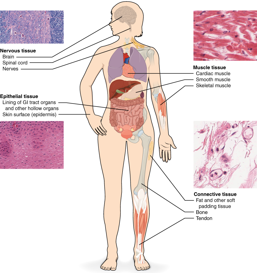
Animals have four types of tissues - connective, epithelial, nervous, and muscle tissue.
1. Connective is made of extracellular matrix-like blood has plasma. Its function is to hold the organs in place. A certain example of the connective is ligament, tendon, bone, adipose and areolar. Ligaments do bone to the bone attachment. Their rigidity will maintain the body structure. Bone store minerals and need calcium for maintaining structure. Further classification of the connective is fibrous, skeletal, and fluid.
2. Epithelial covers the skin surface of organs. They have semi-permeable tight junctions. It will function to provide a barrier between the organ and the external environment. These are specialized to perform excretion, absorption, and secretion. Certain epithelial tissues are simple squamous, simple cuboidal, ciliated, stratified keratinized, stratified non keratinized, etc.
3. The nervous is part of the central nervous system and peripheral nervous system. The CNS includes the spinal cord and brain. The cranial nerves and spinal nerves are part of the peripheral nervous system. They function to maintain the signaling of the transmitters and information from the brain to the body's reflexes.
4. Muscular form the contractile tissue of the body. Its function is to produce motion and force. It is categorized into skeletal, cardiac, and smooth. Cardiac muscle is found in the heart for the contraction of blood throughout the body. Smooth muscle is found in the inner linings of organs. Skeletal attaches to the bone (providing structure, stability, and rigidity), and the function is to generate movement.

Plants have three types of tissue - epidermis, vascular, and ground tissues.
1. The epidermis is the outer layer of tissue present on leaves and young plants, making their structure.
2. Vascular makes the xylem and phloem, which transport fluids and food throughout the plant.
3. The ground makes the nutrients by photosynthesis and makes them store in the leaves. Therefore, this tissue is less differentiated.
The plants have other types of tissue - Meristematic and permanent
1. Meristematic are actively dividing cells which will increase the thickness and length of the plant. They are present at the tips of stems and roots. Certain meristematic tissues are apical meristem, lateral meristem, and intercalary meristem.
2. Permanent cells have taken up the specific function and cannot divide further. They are of 3 types - parenchyma cells, sclerenchyma cells, and collenchyma cells.
Sample Preparation
The success of histology depends on the efficiency of specimen collection and the method of observation.
1. Fixation:
This technique will harden the specimen cells to be observed under the microscope not to move. Fixatives are used to preserve and maintain their structure. The most used fixative is formalin in light microscopy. In electron microscopy, glutaraldehyde, osmium tetroxide is used. The function of fixatives is to cross-link amino groups in protein by forming methylene bridges; this structural integrity of cells and tissues can sometimes lead to damage of proteins like enzymes.
2. Selection and Trimming
The appropriate cell and tissue are needed to be selected and mounted on the microscope. So only the required part is selected and trimmed to be examined. Trimming will cut the sample portion for later sectioning. These samples are made to fit in the cassettes.
3. Embedding
This is done to support the specimen into a harder medium. This is generally done after dehydration. Then the medium is replaced with the solidifying medium and an intermediary fluid which is miscible in embedding media. Like paraffin wax, in light microscopy, it is the most used embedding media. Paraffin wax is immiscible in water, so the water is dehydrated first, and clearing agents are added to remove any alcohol used to remove water. Thus dehydration, clearing, and wax infiltrations are done progressively in histology.
4. Sectioning
A knife is mounted on the microtome to retrieve the sections to be mounted on the slide. The size is usually around 5-15 micrometers. For TEM (Transmission Electron Microscopy), a glass knife cuts 50-175 nanometre thick tissue.
5. Staining
It is the most important part of histology. It is done to give a contrast to the cells which we want to study. We can stain either the background or can stain the specimen cells depending upon the requirement. Histochemistry is when the stain is used to target a specific part of the tissue or cell. Generally, hematoxylin is a commonly used stain for studying the structure of cells and tissues. The stain is directly poured dropwise on the slide with the specimen. It is kept for a few seconds or minutes and is washed off to get the blue stain, not the staining dye's dark color. The slide is then observed under specific conditions to study the stained cells. Histology includes different stains for different techniques used like historiography, electron microscopy, and immunohistochemistry.

5. Specialized Techniques
In histology, frozen sectioning is commonly called cryosectioning, which includes rapid freeze, cut, and mounting of histology samples. Another technique is ultramicrotomy, in which the ultra-thin sample size is obtained for histology under the electron microscope analysis. This process of ultramicrotomy is done by using ultramicrotome to get thin sections of about 0.1 micrometers.
6. Artifacts
These are the structure which can be a hurdle in the histology that is these structures interfere with the observations.
In Vivo Histology
In vivo histology is a new technology being invented that will allow the doctor to directly examine the patients' cells and tissue in a living state. This will eradicate the problem of obtaining a sample and examining them under the microscopes.
Common Mistakes
- The sectioning and mounting have to be very precise.
- Staining should never be overdone.
Context and Applications
This topic is significant in the professional exams for both undergraduate and graduate courses, especially for
- Bachelor of Science in Biology
- Master of Science in Zoology
- Bachelor of Medicine
Related Concepts
- Cytology
- Paleontology
- Histopathology
Want more help with your biology homework?
*Response times may vary by subject and question complexity. Median response time is 34 minutes for paid subscribers and may be longer for promotional offers.
Histology Homework Questions from Fellow Students
Browse our recently answered Histology homework questions.