How does the microscopy in Figure 2 show that the capsule and an S-layer can exist in the same cell at the same time? I need help finding the answer in the article and explain in short answer
How does the microscopy in Figure 2 show that the capsule and an S-layer can exist in the same cell at the same time? I need help finding the answer in the article and explain in short answer
How does the microscopy in Figure 2 show that the capsule and an S-layer can exist in the same cell at the same time? I need help finding the answer in the article and explain in short answer
How does the microscopy in Figure 2 show that the capsule and an S-layer can exist in the same cell at the same time?
I need help finding the answer in the article and explain in short answer
link to article: https://www.ncbi.nlm.nih.gov/pmc/articles/PMC106848/
Transcribed Image Text:54
A
B
MESNAGE ET AL.
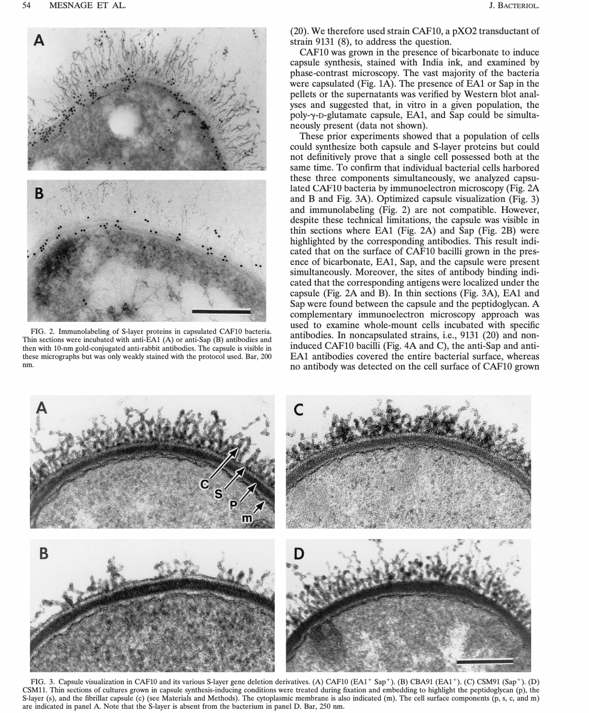
FIG. 2. Immunolabeling of S-layer proteins in capsulated CAF10 bacteria.
Thin sections were incubated with anti-EA1 (A) or anti-Sap (B) antibodies and
then with 10-nm gold-conjugated anti-rabbit antibodies. The capsule is visible in
these micrographs but was only weakly stained with the protocol used. Bar, 200
nm.
A
B
C
m
(20). We therefore used strain CAF10, a pXO2 transductant of
strain 9131 (8), to address the question.
J. BACTERIOL.
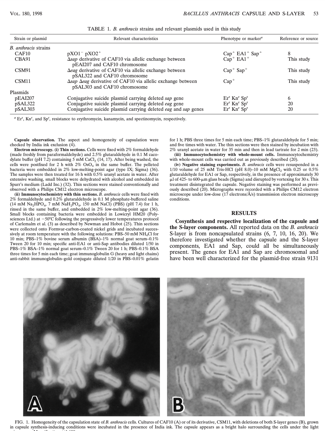
CAF10 was grown in the presence of bicarbonate to induce
capsule synthesis, stained with India ink, and examined by
phase-contrast microscopy. The vast majority of the bacteria
were capsulated (Fig. 1A). The presence of EA1 or Sap in the
pellets or the supernatants was verified by Western blot anal-
yses and suggested that, in vitro in a given population, the
poly-y-D-glutamate capsule, EA1, and Sap could be simulta-
neously present (data not shown).
These prior experiments showed that a population of cells
could synthesize both capsule and S-layer proteins but could
not definitively prove that a single cell possessed both at the
same time. To confirm that individual bacterial cells harbored
these three components simultaneously, we analyzed capsu-
lated CAF10 bacteria by immunoelectron microscopy (Fig. 2A
and B and Fig. 3A). Optimized capsule visualization (Fig. 3)
and immunolabeling (Fig. 2) are not compatible. However,
despite these technical limitations, the capsule was visible in
thin sections where EA1 (Fig. 2A) and Sap (Fig. 2B) were
highlighted by the corresponding antibodies. This result indi-
cated that on the surface of CAF10 bacilli grown in the pres-
ence of bicarbonate, EA1, Sap, and the capsule were present
simultaneously. Moreover, the sites of antibody binding indi-
cated that the corresponding antigens were localized under the
capsule (Fig. 2A and B). In thin sections (Fig. 3A), EA1 and
Sap were found between the capsule and the peptidoglycan. A
complementary immunoelectron microscopy approach was
used to examine whole-mount cells incubated with specific
antibodies. In noncapsulated strains, i.e., 9131 (20) and non-
induced CAF10 bacilli (Fig. 4A and C), the anti-Sap and anti-
EA1 antibodies covered the entire bacterial surface, whereas
no antibody was detected on the cell surface of CAF10 grown
C
D
FIG. 3. Capsule visualization in CAF10 and its various S-layer gene deletion derivatives. (A) CAF10 (EA1+ Sap*). (B) CBA91 (EA1+). (C) CSM91 (Sap+). (D)
CSM11. Thin sections of cultures grown in capsule synthesis-inducing conditions were treated during fixation and embedding to highlight the peptidoglycan (p), the
S-layer (s), and the fibrillar capsule (c) (see Materials and Methods). The cytoplasmic membrane is also indicated (m). The cell surface components (p, s, c, and m)
are indicated in panel A. Note that the S-layer is absent from the bacterium in panel D. Bar, 250 nm.
Transcribed Image Text:VOL. 180, 1998
Strain or plasmid
B. anthracis strains
CAF10
CBA91
CSM91
CSM11
Plasmids
PEAI207
PSAL322
PSAL303
TABLE 1. B. anthracis strains and relevant plasmids used in this study
Relevant characteristics
pX01 PXO2+
Asap derivative of CAF10 via allelic exchange between
PEAI207 and CAF10 chromosome
Aeag derivative of CAF10 via allelic exchange between
pSAL322 and CAF10 chromosome
Asap Aeag derivative of CAF10 via allelic exchange between
pSAL303 and CAF10 chromosome
BACILLUS ANTHRACIS CAPSULE AND S-LAYER 53
Conjugative suicide plasmid carrying deleted sap gene
Conjugative suicide plasmid carrying deleted eag gene
Conjugative suicide plasmid carrying deleted eag and sap genes
"Er', Kn', and Sp', resistance to erythromycin, kanamycin, and spectinomycin, respectively.
Capsule observation. The aspect and homogeneity of capsulation were
checked by India ink exclusion (4).
Electron microscopy. (i) Thin sections. Cells were fixed with 2% formaldehyde
(made freshly from paraformaldehyde) and 2.5% glutaraldehyde in 0.1 M caco-
dylate buffer (pH 7.2) containing 5 mM CaCl₂ (14, 17). After being washed, the
cells were postfixed for 2 h with 2% OsO4 in the same buffer. The pelleted
bacteria were embedded in 2% low-melting-point agar (type IX; Sigma) (36).
The samples were then treated for 16 h with 0.5% uranyl acetate in water. After
extensive washing, small blocks were dehydrated with alcohol and embedded in
Spurr's medium (Ladd Inc.) (32). Thin sections were stained conventionally and
observed with a Philips CM12 electron microscope.
(ii) Immunocytochemistry with thin sections. B. anthracis cells were fixed with
2% formaldehyde and 0.2% glutaraldehyde in 0.1 M phosphate-buffered saline
(14 mM Na₂HPO4, 7 mM NaH₂PO4, 150 mM NaCl) (PBS) (pH 7.4) for 1 h,
rinsed in the same buffer, and embedded in 2% low-melting-point agar (36).
Small blocks containing bacteria were embedded in Lowicryl HM20 (Poly-
sciences Ltd.) at -50°C following the progressively lower temperatures protocol
of Carlemalm et al. (3) as described by Newman and Hobot (25). Thin sections
were collected onto Formvar-carbon-coated nickel grids and incubated succes-
sively at room temperature with the following solutions: PBS-50 mM NH4Cl for
10 min; PBS-1% bovine serum albumin (BSA) -1% normal goat serum-0.1%
Tween 20 for 10 min; specific anti-EA1 or anti-Sap antibodies diluted 1/50 in
PBS-1% BSA-1% normal goat serum-0.1% Tween 20 for 1 h; PBS-0.1% BSA
three times for 5 min each time; goat immunoglobulin G (heavy and light chains)
anti-rabbit immunoglobulin-gold conjugate diluted 1/20 in PBS-0.01% gelatin
Phenotype or marker
Cap EA1 Sap+
Cap+ EA1+
Cap+ Sap+
Cap
Er Kn¹ Sp
Er Kn' Sp
Er Kn¹ Sp¹
Reference or source
RESULTS
8
This study
This study
This study
6
20
20
for 1 h; PBS three times for 5 min each time; PBS-1% glutaraldehyde for 5 min;
and five times with water. The thin sections were then stained by incubation with
2% uranyl acetate in water for 35 min and then in lead tartrate for 2 min (23).
(iii) Immunocytochemistry with whole-mount cells. Immunocytochemistry
with whole-mount cells was carried out as previously described (20).
(iv) Negative staining experiments. B. anthracis cells were resuspended in a
1/10 volume 25 mM Tris-HCl (pH 8.0)-10 mM MgCl₂ with 0.25 or 0.5%
glutaraldehyde for EA1 or Sap, respectively, in the presence of approximately 30
μl of 425- to 600-μm glass beads (Sigma) and disrupted by vortexing for 30 s. This
treatment disintegrated the capsule. Negative staining was performed as previ-
ously described (20). Micrographs were recorded with a Philips CM12 electron
microscope under low-dose (17 electrons/Å/s) transmission electron microscopy
conditions.
Cosynthesis and respective localization of the capsule and
the S-layer components. All reported data on the B. anthracis
S-layer is from noncapsulated strains (6, 7, 10, 16, 20). We
therefore investigated whether the capsule and the S-layer
components, EA1 and Sap, could all be simultaneously
present. The genes for EA1 and Sap are chromosomal and
have been well characterized for the plasmid-free strain 9131
A
B
FIG. 1. Homogeneity of the capsulation state of B. anthracis cells. Cultures of CAF10 (A) or of its derivative, CSM11, with deletions of both S-layer genes (B), grown
in capsule synthesis-inducing conditions were incubated in the presence of India ink. The capsule appears as a bright halo surrounding the cells under the light
Expert Solution
This question has been solved!
Explore an expertly crafted, step-by-step solution for a thorough understanding of key concepts.
Need a deep-dive on the concept behind this application? Look no further. Learn more about this topic, biology and related others by exploring similar questions and additional content below.