L K M J H Table 2. Structures of the Human Eye Label in Structure Name Function figure aqueous humor vitreous humor suspensory ligament sclera N retina pupil optic nerve optic disk (blind spot) lens iris fovea cornea choroid central artery and vein of retina B 6. C D 7. 8. G Functions: 1. Gives the eye its shape; protection. 2. Allows light into the eye; refracts light and helps the eye to focus, along with the lens. 3. Transmits visual information from the eye to the E brain 4. Attach the lens to ciliary muscle; contributes to changing the shape of the lens for focusing 5. Fills the anterior chamber and maintains the shape of the cornea Contains connective tissue and blood vessels that supply nutrients to the retina and remove wastes from the retina Opening that lets light into the eye Controls the size of pupil to let more or less light into the eve 9. Fills the posterior chamber and helps the eyeball maintain shape. 10. Contains photoreceptor cells that respond to light and initiate signals to the brain. 11. The area where the optic nerve passes through the retina. 12. Focuses light on the retina. 13. Contains the highest concentration of photoreceptor cells; provides the sharpest focus as the center of the visual field. 14. Supplies oxygen and nutrients to choroid and retina; removes waste therefrom
L K M J H Table 2. Structures of the Human Eye Label in Structure Name Function figure aqueous humor vitreous humor suspensory ligament sclera N retina pupil optic nerve optic disk (blind spot) lens iris fovea cornea choroid central artery and vein of retina B 6. C D 7. 8. G Functions: 1. Gives the eye its shape; protection. 2. Allows light into the eye; refracts light and helps the eye to focus, along with the lens. 3. Transmits visual information from the eye to the E brain 4. Attach the lens to ciliary muscle; contributes to changing the shape of the lens for focusing 5. Fills the anterior chamber and maintains the shape of the cornea Contains connective tissue and blood vessels that supply nutrients to the retina and remove wastes from the retina Opening that lets light into the eye Controls the size of pupil to let more or less light into the eve 9. Fills the posterior chamber and helps the eyeball maintain shape. 10. Contains photoreceptor cells that respond to light and initiate signals to the brain. 11. The area where the optic nerve passes through the retina. 12. Focuses light on the retina. 13. Contains the highest concentration of photoreceptor cells; provides the sharpest focus as the center of the visual field. 14. Supplies oxygen and nutrients to choroid and retina; removes waste therefrom

Human Anatomy & Physiology (11th Edition)
11th Edition
ISBN:9780134580999
Author:Elaine N. Marieb, Katja N. Hoehn
Publisher:Elaine N. Marieb, Katja N. Hoehn
Chapter1: The Human Body: An Orientation
Section: Chapter Questions
Problem 1RQ: The correct sequence of levels forming the structural hierarchy is A. (a) organ, organ system,...

Related questions
Question
Transcribed Image Text:L
A
K
I
M
J
H.
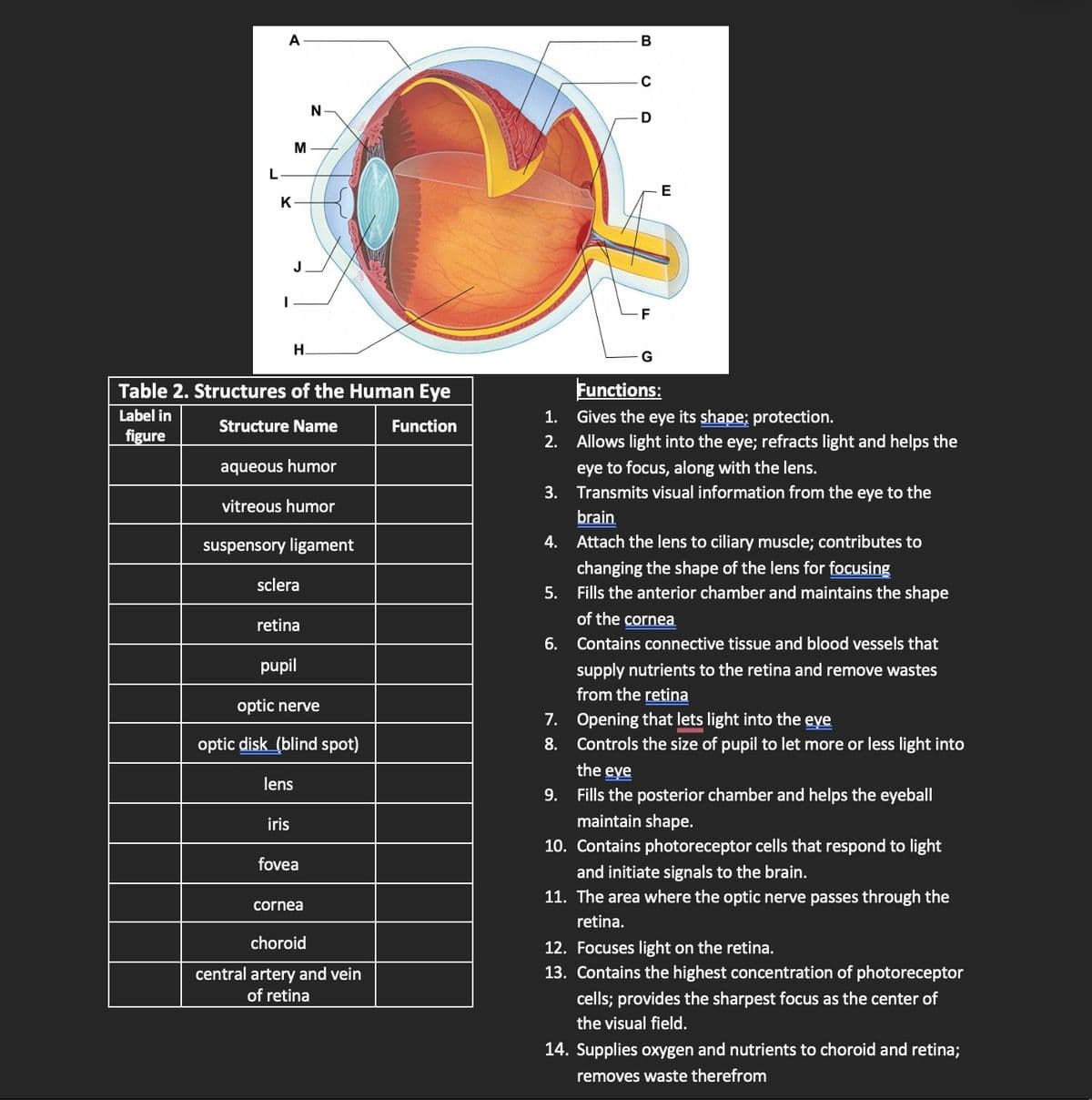
Table 2. Structures of the Human Eye
Label in
Structure Name
Function
figure
aqueous humor
vitreous humor
suspensory ligament
sclera
retina
pupil
iris
N
optic nerve
optic disk (blind spot)
lens
fovea
cornea
choroid
central artery and vein
of retina
1.
2.
B
C
7.
8.
D
-F
E
Functions:
Gives the eye its shape; protection.
Allows light into the eye; refracts light and helps the
eye to focus, along with the lens.
3.
Transmits visual information from the eye to the
brain
4.
Attach the lens to ciliary muscle; contributes to
changing the shape of the lens for focusing
5. Fills the anterior chamber and maintains the shape
of the cornea
6. Contains connective tissue and blood vessels that
supply nutrients to the retina and remove wastes
from the retina
Opening that lets light into the eve
Controls the size of pupil to let more or less light into
the eve
9.
Fills the posterior chamber and helps the eyeball
maintain shape.
10. Contains photoreceptor cells that respond to light
and initiate signals to the brain.
11. The area where the optic nerve passes through the
retina.
12. Focuses light on the retina.
13. Contains the highest concentration of photoreceptor
cells; provides the sharpest focus as the center of
the visual field.
14. Supplies oxygen and nutrients to choroid and retina;
removes waste therefrom
Expert Solution
This question has been solved!
Explore an expertly crafted, step-by-step solution for a thorough understanding of key concepts.

This is a popular solution!

Trending now
This is a popular solution!

Step by step
Solved in 3 steps

Recommended textbooks for you
Human Anatomy & Physiology (11th Edition)
Anatomy and Physiology
ISBN:
9780134580999
Author:
Elaine N. Marieb, Katja N. Hoehn
Publisher:
PEARSON

Anatomy & Physiology
Anatomy and Physiology
ISBN:
9781259398629
Author:
McKinley, Michael P., O'loughlin, Valerie Dean, Bidle, Theresa Stouter
Publisher:
Mcgraw Hill Education,

Human Anatomy
Anatomy and Physiology
ISBN:
9780135168059
Author:
Marieb, Elaine Nicpon, Brady, Patricia, Mallatt, Jon
Publisher:
Pearson Education, Inc.,
Human Anatomy & Physiology (11th Edition)
Anatomy and Physiology
ISBN:
9780134580999
Author:
Elaine N. Marieb, Katja N. Hoehn
Publisher:
PEARSON
Anatomy & Physiology
Anatomy and Physiology
ISBN:
9781259398629
Author:
McKinley, Michael P., O'loughlin, Valerie Dean, Bidle, Theresa Stouter
Publisher:
Mcgraw Hill Education,
Human Anatomy
Anatomy and Physiology
ISBN:
9780135168059
Author:
Marieb, Elaine Nicpon, Brady, Patricia, Mallatt, Jon
Publisher:
Pearson Education, Inc.,

Anatomy & Physiology: An Integrative Approach
Anatomy and Physiology
ISBN:
9780078024283
Author:
Michael McKinley Dr., Valerie O'Loughlin, Theresa Bidle
Publisher:
McGraw-Hill Education

Human Anatomy & Physiology (Marieb, Human Anatomy…
Anatomy and Physiology
ISBN:
9780321927040
Author:
Elaine N. Marieb, Katja Hoehn
Publisher:
PEARSON