Movement of the Specimen on the Stage and its Corresponding Movement in the eyepiece: Table 2-Movement of specimens If letter e on stage is moved to: The image in the eyepiece is moved to: 3 o'clock o'clock 9 o'clock o'clock 12 o'clock o'clock 6 o'clock o'clock
Movement of the Specimen on the Stage and its Corresponding Movement in the eyepiece: Table 2-Movement of specimens If letter e on stage is moved to: The image in the eyepiece is moved to: 3 o'clock o'clock 9 o'clock o'clock 12 o'clock o'clock 6 o'clock o'clock

Biomedical Instrumentation Systems
1st Edition
ISBN:9781133478294
Author:Chatterjee
Publisher:Chatterjee
Chapter16: Fiber Optics And Lasers In Bioinstrumentation
Section: Chapter Questions
Problem 5Q

Related questions
Question
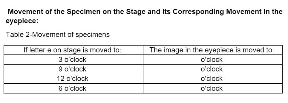
Transcribed Image Text:Movement of the Specimen on the Stage and its Corresponding Movement in the
eyepiece:
Table 2-Movement of specimens
If letter e on stage is moved to:
The image in the eyepiece is moved to:
3 o'clock
o'clock
9 o'clock
o'clock
12 o'clock
o'clock
6 o'clock
o'clock
Expert Solution
This question has been solved!
Explore an expertly crafted, step-by-step solution for a thorough understanding of key concepts.

This is a popular solution!

Trending now
This is a popular solution!

Step by step
Solved in 2 steps

Recommended textbooks for you

Principles Of Radiographic Imaging: An Art And A …
Health & Nutrition
ISBN:
9781337711067
Author:
Richard R. Carlton, Arlene M. Adler, Vesna Balac
Publisher:
Cengage Learning
Principles Of Radiographic Imaging: An Art And A …
Health & Nutrition
ISBN:
9781337711067
Author:
Richard R. Carlton, Arlene M. Adler, Vesna Balac
Publisher:
Cengage Learning