Table 2 – Movement of specimens If the letter e on stage Is moved to 3 o'clock The image in the eyepiece is moved to: O'clock 9 o'clock O'clock O'clock O'clock 12 o'clock 6 o'clock
Table 2 – Movement of specimens If the letter e on stage Is moved to 3 o'clock The image in the eyepiece is moved to: O'clock 9 o'clock O'clock O'clock O'clock 12 o'clock 6 o'clock

Chapter3: Tools For Exploring The World: Physical, Perceptual, And Motor Development
Section3.4: Coming To Know The World
Problem 5TY

Related questions
Question
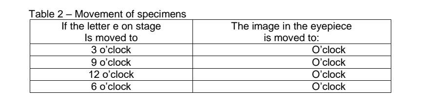
Transcribed Image Text:Table 2 – Movement of specimens
If the letter e on stage
Is moved to
The image in the eyepiece
is moved to:
3 o'clock
O'clock
9 o'clock
12 o'clock
6 o'clock
O'clock
O'clock
O'clock
Expert Solution
This question has been solved!
Explore an expertly crafted, step-by-step solution for a thorough understanding of key concepts.

Step by step
Solved in 2 steps

Recommended textbooks for you

Principles Of Radiographic Imaging: An Art And A …
Health & Nutrition
ISBN:
9781337711067
Author:
Richard R. Carlton, Arlene M. Adler, Vesna Balac
Publisher:
Cengage Learning

Biology 2e
Biology
ISBN:
9781947172517
Author:
Matthew Douglas, Jung Choi, Mary Ann Clark
Publisher:
OpenStax
Principles Of Radiographic Imaging: An Art And A …
Health & Nutrition
ISBN:
9781337711067
Author:
Richard R. Carlton, Arlene M. Adler, Vesna Balac
Publisher:
Cengage Learning
Biology 2e
Biology
ISBN:
9781947172517
Author:
Matthew Douglas, Jung Choi, Mary Ann Clark
Publisher:
OpenStax

Anatomy & Physiology
Biology
ISBN:
9781938168130
Author:
Kelly A. Young, James A. Wise, Peter DeSaix, Dean H. Kruse, Brandon Poe, Eddie Johnson, Jody E. Johnson, Oksana Korol, J. Gordon Betts, Mark Womble
Publisher:
OpenStax College