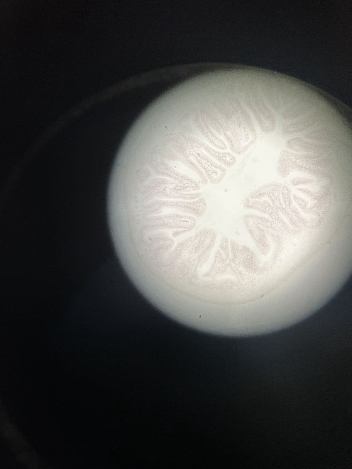
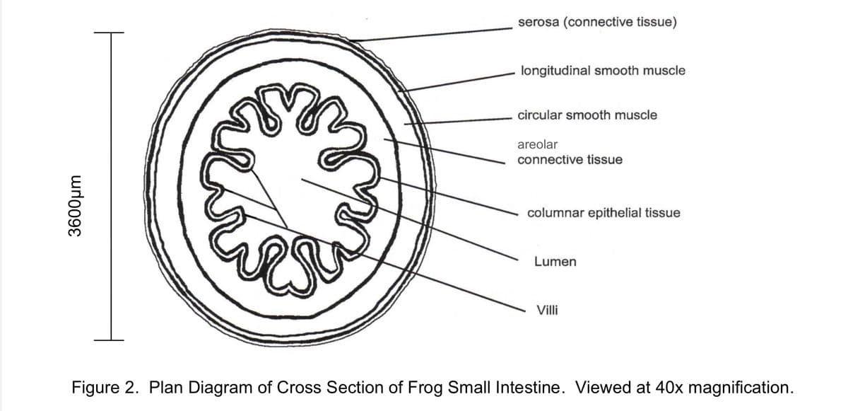
Objective: Drawing a plan diagram of a frog intensine viewed under a microscope, as well as calculating actual size and drawing magnification of the width of the intestine. Please see the attached microscopic image of the frog intestine when viewed under the microscope, and an example labeled drawing of the intestine. I approximated that 3-4 cells would fit across (frog intenstine is much smaller when viewed under microscope, photo taken appears much longer). 1. Draw a plan diagram and label the following: • Longitudinal smooth muscle • Columnar epithelial tissue • Circular smooth muscle • Villi • Areolar connective tissue • Lumen 2. Calculate actual size and drawing magnification of the width of the intestine. Include formula used and calculations. 2. Add a scale bar with actual size next to the diagram. 3. Give the drawing a descriptive title and record total magnification. Thank you!


Objective: Drawing a plan diagram of a frog intensine viewed under a microscope, as well as calculating actual size and drawing magnification of the width of the intestine.
Please see the attached microscopic image of the frog intestine when viewed under the microscope, and an example labeled drawing of the intestine. I approximated that 3-4 cells would fit across (frog intenstine is much smaller when viewed under microscope, photo taken appears much longer).
1. Draw a plan diagram and label the following: • Longitudinal smooth muscle • Columnar epithelial tissue • Circular smooth muscle • Villi • Areolar connective tissue • Lumen
2. Calculate actual size and drawing magnification of the width of the intestine. Include formula used and calculations.
2. Add a scale bar with actual size next to the diagram.
3. Give the drawing a descriptive title and record total magnification.
Thank you!


Trending now
This is a popular solution!

Step by step
Solved in 3 steps


