Which of the following is/are TRUE regarding microscopes? * TEM gives excellent view of internal structures of a specimen in three- dimensional image Phase contrast microscope permits detailed examination of internal structures of live specimens Interference microscope creates two superimposed images of a specimen.
Q: Differentiate between the limit of resolution of the typical light microscope and that of the…
A: The microscope is an instrument that helps us to visualize the objects that cannot be seen by our…
Q: 9. 2. 10. 4. 11. 5. 6. 12. 7. 13. 8. 14. LI. 3. 5.
A: the given figure represnts a simple microscope , used to magnify small objects and specimens . used…
Q: 
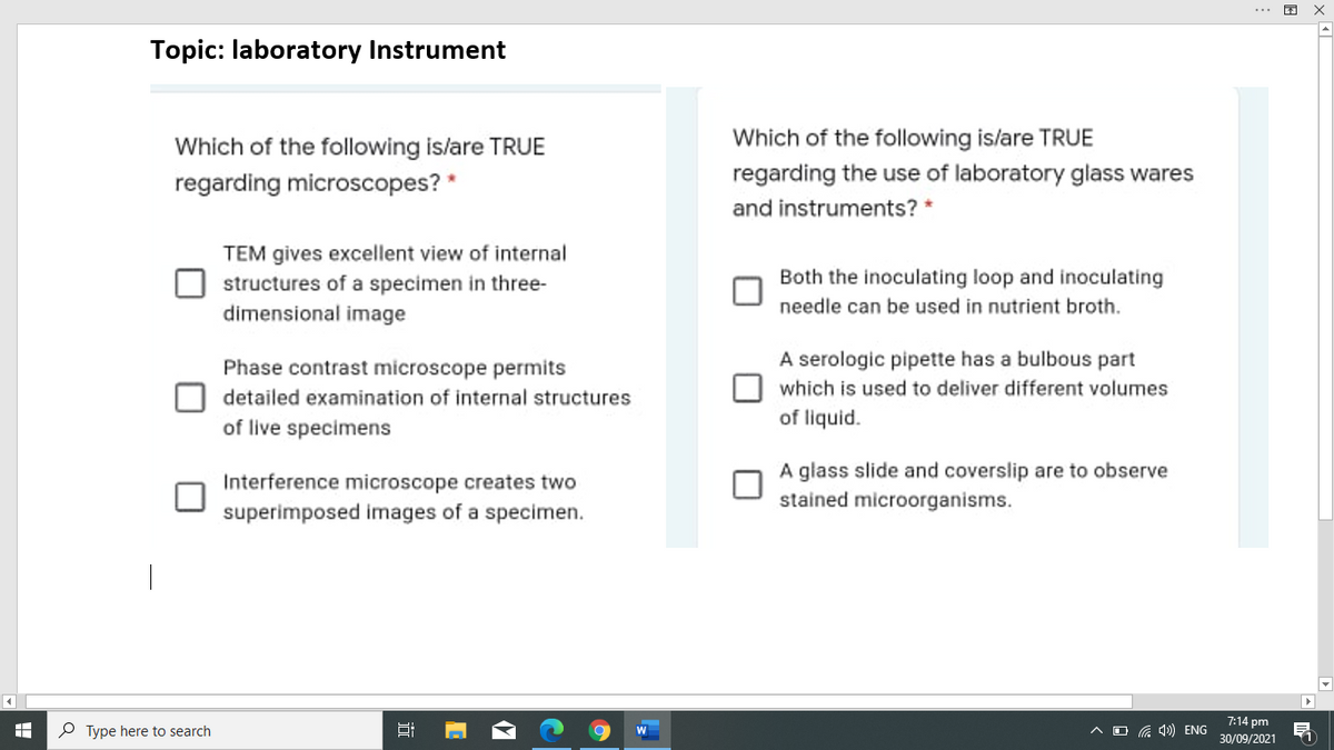
Topic: laboratory Instrument Which of the following is/are TRUE Which of the following is/are TRUE…
A: Microscope are the instrument used to view enlarged image of a minute structures at a scale…
Q: Which of the following is never useful for observing living cells? O phase-contrast microscope…
A: Introduction All living organisms are made up of cells, which are the basic building components.…
Q: Match the terms on the left with its description or definition on the right. There is only one…
A: (Please note you have posted multiple questions. We can solve 1 question at a time. Please post rest…
Q: Both light and electron microscopy are commonly used to visualize cells, cell structures, and the…
A: The cell is the basic structural and functional unit of the body. It is very important to determine…
Q: All of the following are types of light microscopes except electron phase-contrast Obright-field O…
A: Introduction Micoscope is a laboratory instrument. It is used to observe very small object which can…
Q: Which of the following microscopes typically requires the use of vital dyes (like methylene blue) to…
A: Microscopy is the technical field of using microscopes to view objects and areas of objects that…
Q: discuss the microscopy technique that gives the best constrast image of non biological samples such…
A: Microscopy is an instrument used to magnify the size of the objects or particle which cannot be seen…
Q: Confocal microscopy uses laser light to control the depth of field, and allowing visualisation of an…
A: Microscopy refers to the study of minute structures and objects by the use of an instrument called…
Q: WHAT ARE THE USES OF THE FOLLOWING MICROSCOPE AND THEIR RELEVANCE IN THE MEDICAL FIELD? -COMPOUND…
A: Microscope is an equipment that enable us to see magnified version of minute specimen and objects.…
Q: Compare the appearance of microorganisms you observed using phase-contrast microscopy versus…
A: Microscopy is a basic form of microscopic technology. It depends on the interaction between light…
Q: Compare the 2 types of Bright-field microscopes. Why is a microscope is an important tool in the…
A: A microscope is a scientific tool that is used to look at items that are too small to view with the…
Q: 1. Differentiate the compound light microscope from the dissecting microscope in terms of: *image…
A: Microscopy is a technique of incorporating microscopes to enlarge the size of objects (invisible to…
Q: electron microscope It is a special type of microscope having a high resolution of images, able to…
A: Electron microscope Electron microscope utilise is electrons as a source of illumination for…
Q: Rank the three main types of microscopes from lowest to highestpotential magnification.
A: Magnification is measure of the ability of a lens or other optical instruments to magnify, expressed…
Q: What is the magnification of the microscope shown below? Grid your answer. 10x O OG
A: Microscopes are instruments used to examine objects that are not seen through the naked eye. They…
Q: Compare and contrast the optical compound microscope with theelectron microscope.
A: An optical Compound microscope is the simplest microscope. An electron microscope is a complex…
Q: Two monocular compound microscopes properly spacedand aligned describe the__________ microscope.
A: Stereoscopic/Stereo/Dissecting microscope It is an optical microscope varient designed for low…
Q: Microscope Structure and Function Compound Light Microscope Structures: Label the names of the…
A: Microscope can be defined as the instrument which is used to see the organisms or objects that is…
Q: Which of the following microscopes allows study of living specimens? The scanning electron…
A: Microscopy is the branch of biological sciences in which the minute structures like the cells and…
Q: WHAT ARE THE DIFFERENT TYPES OF MICROSCOPES AND THEIR USES?
A: Microscope is the widely used laboratory instrument in the molecular biology, it is mainly used to…
Q: Hi, there! Just subscribed to Bartleby! So excited to be here, and thanks for your help! Can you…
A: An instrument that is used to see extremely small objects that cannot be seen by naked eyes is…
Q: Which of the following statements is correct about the difference between dissecting microscope and…
A: Dissecting Microscope - The dissecting microscope has the stage has the stage below the light…
Q: TEST I. Fill in the necessary concepts about microscopes. OF COMPounb dissection or stereoscope…
A: Dissection microscope is also known as simple microscope. It has only one lens. Compound microscope…
Q: Fill in table 1 below for the characteristics of your microscopes. Table 1- Characteristics of the…
A: Light microscopes work on a combination of lenses that magnify the image of the object making it…
Q: How do the different parts of microscope serve their specific function? Cite examples
A: Introduction Cell is the basic unit of life which perform all kind of physiological and metabolic…
Q: ical Microscopy Label the parts & functions of a COMPOUND LIGHT MICROSCOPE How to set-up a…
A: Functions of a microscope : The light microscope is an instrument for visualizing fine detail of an…
Q: What are the mechanical parts of microscope and their functions?
A: Many important anatomical features, particularly those that function at the tissue or cellular…
Q: All of the following are types of light microscopes EXCEPT O fluorescent. O scanning tunneling. O…
A: Light microscopy Light microscopy is also called ptical microscopy, which uses a visible light…
Q: The above photograph (IMAGE ATTACHED) of a cheek smear was obtained using the high-power objective…
A: The eyepiece graticule is a scale that is placed in the eyepiece of a microscope containing scales…
Q: A microscope is turned on, under low power, with a slide positioned on the stage ready to focus.…
A: A microscope is an important tool that is used to view microscopic specimens. There are many types…
Q: What are microscopes?
A: Microscope invention is a revolution for mankind to study minute living and nonliving things. It…
Q: Describe the operating features of electron microscopes andhow they differ from optical microscopes…
A: Optical microscopes use light as a source for image formation. Unlike the light or optical…
Q: Which of the following microscopes allows study of living specimens? The light compound microscope O…
A: Microscopy is the branch of biological sciences in which the structures that are usually not visible…
Q: Make a diagram of the transmission electron microscope with all its parts and describe them
A: The microscope is a device that allows you to magnify small objects. A microscope can be used to…
Q: List the different parts of the microscope, gives its classification and function.
A: Microscopes are the instruments which are routinely used in labs to visualised very small objects…
Q: Drag the images or descriptions to their corresponding class to test your understanding of various…
A: Microscope The device which is use to visualise microorganisms which are not visible to the naked…
Q: The use of the natural source of light collected by the mirror and condensed by the diaphragm is a…
A: Microscope, a tool that magnifies pictures of small objects, enabling the spectator to have an…
Q: TRUE or FALSE only! 1. Microscopes are necessary for biological investigations because it allows us…
A: Since you have posted a question with multiple sub-parts, we will solve the first three sub-parts…
Q: Non-conducting materials can be studied by scanning tunneling microscope(STM). 1.True 2. False
A: INTRODUCTION Scanning and tunneling microscope (STM) STM is a kind of microscope that is used to…
Q: The resolving power of the microscope reveals: a. Whether or not oil is needed to see a…
A: The instrument that is utilized in the laboratories to visualize the objects or structures that are…
Q: Describe 3 simple ways of preparing slides for light microscopy microscope
A: Introduction The optical microscope, often known as a light microscope, is a type of microscope that…
Q: You are trying to look at a specimen with a compound microscope using the 4x objective, but it is…
A: A microscope is an instrument that amplifies an article so it might be seen by the eyewitness. Since…
Q: discuss the use and applications of MICROSCOPE in microbiology and clinical studies. Parts and…
A: Microorganisms (Bacteria, viruses, fungi) are tiny living cells that are too small to see through…
Q: 5 1 2 6. 4 7 8.
A: Introduction A microscope is a scientific tool that is used to study items that are too small for…
Trending now
This is a popular solution!
Step by step
Solved in 3 steps
- discuss the microscopy technique that gives the best constrast image of non biological samples such as micro and nano particles?Student Y is working with his microbiology experiment, the directions of the agar is to suspend 25g in 1000mL of distilled water, heat to boiling until completely dissolve. How much of the agar does he need to prepare for 18 petri dishes?Discuss the microscopy technique that gives the best contrast image of non-biological samples such as micro and nano-particles.
- a. How was the specimen prepared for the microscopy technique applied? (for e.g. stained with H&E stain, Gram stain, unstained) b. What is the microscopy technique and magnification used to obtain this image? c. What is the basic principle of image formation using this microscopy technique? d. What can be observed and concluded from the image of the specimen? e. Are there any potential aberrations present in this specimen image? Describe these and how they may affect interpretation of the result.Describe the operating features of electron microscopes andhow they differ from optical microscopes in illumination source,magnification, resolution, and image appearance.discuss the use and applications of MICROSCOPE in microbiology and clinical studies. Parts and functions. Cite application of microscopy in the discussion
- Explain in detail the study of microbial structure microscopy.Compare the appearance of microorganisms you observed using phase-contrast microscopy versus brightfield microscopy.Which microscopy technique is BEST at determining the presence of physical microbial interaction(s)? a Light microscopy. b Scanning or transmission electron microscopy. c Fluorescent microscopy. d Phase contrast microscopy.
- 1. Differentiate the compound light microscope from the dissecting microscope in terms of: *image produced *magnification *type of specimen examined 2. Write down the principles of the following: *electron microscopy *scanning tunneling microscopy *atomic force microscopyDifferentiate Brownian movement from true motility Methods: Place the prepared specimen under the microscope. Observe for Brownian movement (Non-motile organisms are tiny particles in suspension. Also, they exhibit movement characterized by an oscillating, quivering motion.) Or and True motility (Motility of microorganisms is an independent, true movement of translation brought about by organs of locomotion or by gliding.) With the presented specimen, observe and determine whether the specimen shows true motility or brownian movement.EXPERIMENT 2 Title: Aseptic technique Objectives: To apply the aseptic technique To observe the growth found on the petri dish. Material/apparatus: Gloves, incubator, soap for handwashing, tissues, nutrient agar, 70% alcohol Procedure: Wipe off the bench with 70% alcohol. Draw a line down the middle of the petri dish to divide the plate in half. Label each halves with A and B. Press your unwashed thumb onto the agar at column A. Apply proper handwashing technique. Put the same thumb after washing onto agar at the column B. Incubate the petri dish at 37°C for 24 hours or overnight. Observe and note down the colonies. Results: