Why test that mice infected with B. anthracis produce antibodies to the S-layer proteins? What is the point, what does it tell us? (figure 6) I need help finding the answer in the article and answer as short a possible link to
Why test that mice infected with B. anthracis produce antibodies to the S-layer proteins? What is the point, what does it tell us? (figure 6) I need help finding the answer in the article and answer as short a possible link to

Biology 2e
2nd Edition
ISBN:9781947172517
Author:Matthew Douglas, Jung Choi, Mary Ann Clark
Publisher:Matthew Douglas, Jung Choi, Mary Ann Clark
Chapter22: Prokaryotes: Bacteria And Archaea
Section: Chapter Questions
Problem 24RQ: A person in England arrives at a medical clinic with a fever and swollen lymph nodes shortly after...

Related questions
Question
Why test that mice infected with B. anthracis produce antibodies to the S-layer proteins? What is the point, what does it tell us? (figure 6)
I need help finding the answer in the article and answer as short a possible
link to article: https://www.ncbi.nlm.nih.gov/pmc/articles/PMC106848/
Transcribed Image Text:The Capsule and S-Layer: Two Independent and Yet
Compatible Macromolecular Structures in Bacillus anthracis
STÉPHANE MESNAGE,¹ EVELYNE TOSI-COUTURE,² PIERRE GOUNON,² MICHÈLE MOCK,¹
AND AGNES FOUET¹*
Toxines et Pathogénie Bactériennes (CNRS URA 1858)¹ and Station Centrale de Microscopie Electronique, ²
Institut Pasteur, Paris, France
Received 5 September 1997/Accepted 22 October 1997
Bacillus anthracis, the etiological agent of anthrax, is a gram-positive spore-forming bacterium. Fully virulent
bacilli are toxinogenic and capsulated. Two abundant surface proteins, including the major antigen, are
components of the B. anthracis surface layer (S-layer). The B. anthracis paracrystalline S-layer has previously
only been found in noncapsulated vegetative cells. Here we report that the S-layer proteins are also synthesized
under conditions where the poly-y-D-glutamic acid capsule is present. Structural and immunological analyses
show that the capsule is exterior to and completely covers the S-layer proteins. Nevertheless, analysis of single
and double S-layer protein mutants shows that the presence of these proteins is not required for normal
capsulation of the bacilli. Similarly, the S-layer proteins assemble as a two-dimensional crystal, even in the
presence of the capsule. Thus, both structures are compatible, and yet neither is required for the correct
formation of the other.
Bacillus anthracis, a gram-positive spore-forming bacterium,
is the causative agent of anthrax. This disease, to which many
animals, including humans, are susceptible, involves toxemia
and septicemia. In the mammalian host, B. anthracis bacilli
synthesize two toxins (lethal and edema toxins) (31) and a
capsule (18) encoded by two large plasmids, pX01 and pXO2,
respectively (12, 21). The capsule is composed of poly-y-D-
glutamic acid and has antiphagocytic properties (13, 31, 37).
Although unusual, a similar capsule is also found on Bacillus
licheniformis bacilli (9). In the absence of pXO2 or the inducer
bicarbonate, the cell does not produce a capsule and the cell
wall appears layered. These layers are composed of fragments
displaying a highly patterned ultrastructure (10, 16). This type
of cell surface is now referred to as the surface layer (S-layer).
S-layers are present on the surfaces of many archaea and
bacteria (for reviews, see references 29 and 30). Most are
formed by noncovalent, entropy-driven assembly of a single
(glyco)protein protomer on the bacterial surface, giving rise to
proteinaceous paracrystalline layers. Generally, a single S-
layer is present, constituting 5 to 10% of total cell protein. Its
synthesis is thus presumably energy consuming for the bacte-
rium. Numerous bacteria have S-layers, suggesting that they
play important roles in the interaction between the cell and its
environment. Various functions have been proposed for S-
layers, including shape maintenance and molecular sieving,
and they can serve in phage fixation. The S-layer may be a
virulence factor, protecting pathogenic bacteria against com-
plement killing, facilitating binding of bacteria to host mole-
cules, or enhancing their ability to associate with macrophages
(for reviews, see references 27 and 29).
Some bacteria, such as cyanobacteria or Azotobacter spp.,
possess both a capsule and an S-layer; however, to our knowl-
edge, their structural relationships have not been analyzed
through simultaneous genetic and cytologic studies. Both of
these features have been independently described for the sur-
* Corresponding author. Mailing address: Toxines et Pathogénie
Bactériennes, Institut Pasteur, 28, rue du Dr Roux, 75724 Paris Cedex
15, France. Phone: 33 1 45 68 86 54. Fax: 33 1 45 68 89 54. E-mail:
afouet@pasteur.fr.
52
face of the pathogenic bacterium B. anthracis. The components
of the B. anthracis S-layer are two abundant surface proteins,
EA1 and Sap (6, 20). Previous analyses of the B. anthracis
S-layer used plasmid-cured strains; consequently, the interac-
tion, if any, between the capsule and the S-layer could not be
studied. Temporal or environmental regulation could be such
that only one or the other structure is ever present at the cell
surface. However, we show that S-layer proteins are synthe-
sized under conditions where the bacilli are capsulated. We
determined the localizations of capsule and S-layer compo-
nents and analyzed whether the S-layer is necessary for proper
capsulation. Finally, the assembly of the S-layer proteins in a
two-dimensional crystal was examined in the presence of the
capsule.
MATERIALS AND METHODS
Plasmids, bacterial strains, mating experiments, and culture conditions. The
plasmids used to disrupt sap (encoding Sap), eag (encoding EA1), and both
genes, i.e., PEA1207, pSAL322, and pSAL303, respectively, were described pre-
viously (6, 20) and are listed in Table 1. The construction of B. anthracis CAF10,
a pXO2 transductant of plasmidless strain 9131, and its regulation of capsule
synthesis have already been reported (8). Escherichia coli JM83 harboring
pRK24 was used for mating experiments (34, 35). Allelic exchange was carried
out as previously described (26) with the spectinomycin resistance cassette as a
selectable marker (24). sap, eag, and both genes were disrupted in CAF10 by
heterogramic conjugation, giving CBA91, CSM91, and CSM11, respectively (Ta-
ble 1). E. coli cells were grown in Luria broth on agar plates (22). Capsule
synthesis was induced by growing B. anthracis cells in brain heart infusion me-
dium (Difco Laboratories) in the presence of 0.6% sodium bicarbonate or on
CAP plates (28) in a 5% CO₂ atmosphere for electron microscopy. Antibiotics
were used at the following concentrations: kanamycin, 40 µg/ml for E. coli;
erythromycin, 5 µg/ml for B. anthracis; and spectinomycin, 60 µg/ml for both E.
coli and B. anthracis.
you w
Protein analysis. To test the in vivo expression of EA1 and Sap, the synthesis
of antibodies was assayed. The rationale of this experiment is that antibodies are
produced only if the antigen is synthesized in vivo. Seven Swiss mice were
injected with 106 spores of strain CAF10 and sacrificed after 30 days. Their sera
were pooled. The gel loading samples were obtained as follows. B. anthracis cells
were harvested at an
an optical density at 600 nm of =2. Pellets were washed in 25
mM Tris-HCl (pH 8.0), sonicated until complete clarification, and resuspended
in Laemmli buffer (19). Samples (3 μg of
of pellet protein and 20 μl of trichloro-
acetic acid-precipitated supernatant protein) were loaded on sodium dodecyl
sulfate-10% polyacrylamide gels. Separated proteins were transferred to nitro-
cellulose sheets by use of the Bio-Rad Trans-Blot system. The sera were used at
1/200 dilution. Western blots were developed with the ECL Western blotting
analysis system (Amersham), with a 1/10,000 dilution of the second antibody.
Transcribed Image Text:VOL. 180, 1998
94
67
43
30
20-
1 2
3 4
5 6 7 8
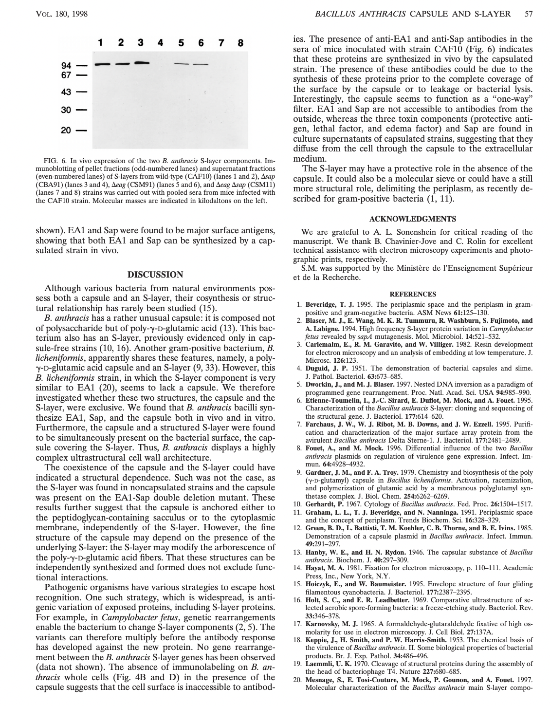
FIG. 6. In vivo expression of the two B. anthracis S-layer components. Im-
munoblotting of pellet fractions (odd-numbered lanes) and supernatant fractions
(even-numbered lanes) of S-layers from wild-type (CAF10) (lanes 1 and 2), Asap
(CBA91) (lanes 3 and 4), Aeag (CSM91) (lanes 5 and 6), and Aeag Asap (CSM11)
(lanes 7 and 8) strains was carried out with pooled sera from mice infected with
the CAF10 strain. Molecular masses are indicated in kilodaltons on the left.
shown). EA1 and Sap were found to be major surface antigens,
showing that both EA1 and Sap can be synthesized by a cap-
sulated strain in vivo.
DISCUSSION
Although various bacteria from natural environments pos-
sess both a capsule and an S-layer, their cosynthesis or struc-
tural relationship has rarely been studied (15).
B. anthracis has a rather unusual capsule: it is composed not
of polysaccharide but of poly-y-D-glutamic acid (13). This bac-
terium also has an S-layer, previously evidenced only in cap-
sule-free strains (10, 16). Another gram-positive bacterium, B.
licheniformis, apparently shares these features, namely, a poly-
y-D-glutamic acid capsule and an S-layer (9, 33). However, this
B. licheniformis strain, in which the S-layer component is very
similar to EA1 (20), seems to lack a capsule. We therefore
investigated whether these two structures, the capsule and the
S-layer, were exclusive. We found that B. anthracis bacilli syn-
thesize EA1, Sap, and the capsule both in vivo and in vitro.
Furthermore, the capsule and a structured S-layer were found
to be simultaneously present on the bacterial surface, the cap-
sule covering the S-layer. Thus, B. anthracis displays a highly
complex ultrastructural cell wall architecture.
The coexistence of the capsule and the S-layer could have
indicated a structural dependence. Such was not the case, as
the S-layer was found in noncapsulated strains and the capsule
was present on the EA1-Sap double deletion mutant. These
results further suggest that the capsule is anchored either to
the peptidoglycan-containing sacculus or to the cytoplasmic
membrane, independently of the S-layer. However, the fine
structure of the capsule may depend on the presence of the
underlying S-layer: the S-layer may modify the arborescence of
the poly-y-D-glutamic acid fibers. That these structures can be
independently synthesized and formed does not exclude func-
tional interactions.
Pathogenic organisms have various strategies to escape host
recognition. One such strategy, which is widespread, is anti-
genic variation of exposed proteins, including S-layer proteins.
For example, in Campylobacter fetus, genetic rearrangements
enable the bacterium to change S-layer components (2, 5). The
variants can therefore multiply before the antibody response
has developed against the new protein. No gene rearrange-
ment between the B. anthracis S-layer genes has been observed
(data not shown). The absence of immunolabeling on B. an-
thracis whole cells (Fig. 4B and D) in the presence of the
capsule suggests that the cell surface is inaccessible to antibod-
BACILLUS ANTHRACIS CAPSULE AND S-LAYER 57
ies. The presence of anti-EA1 and anti-Sap antibodies in the
sera of mice inoculated with strain CAF10 (Fig. 6) indicates
that these proteins are synthesized in vivo by the capsulated
strain. The presence of these antibodies could be due to the
synthesis of these proteins prior to the complete coverage of
the surface by the capsule or to leakage or bacterial lysis.
Interestingly, the capsule seems to function as a "one-way"
filter. EA1 and Sap are not accessible to antibodies from the
outside, whereas the three toxin components (protective anti-
gen, lethal factor, and edema factor) and Sap are found in
culture supernatants of capsulated strains, suggesting that they
diffuse from the cell through the capsule to the extracellular
medium.
The S-layer may have a protective role in the absence of the
capsule. It could also be a molecular sieve or could have a still
more structural role, delimiting the periplasm, as recently de-
scribed for gram-positive bacteria (1, 11).
ACKNOWLEDGMENTS
We are grateful to A. L. Sonenshein for critical reading of the
manuscript. We thank B. Chavinier-Jove and C. Rolin for excellent
technical assistance with electron microscopy experiments and photo-
graphic prints, respectively.
S.M. was supported by the Ministère de l'Enseignement Supérieur
et de la Recherche.
REFERENCES
1. Beveridge, T. J. 1995. The periplasmic space and the periplasm in gram-
positive and gram-negative bacteria. ASM News 61:125-130.
2. Blaser, M. J., E. Wang, M. K. R. Tummuru, R. Washburn, S. Fujimoto, and
A. .Labigne. 1994. High frequency S-layer protein variation in Campylobacter
fetus revealed by sapA mutagenesis. Mol. Microbiol. 14:521-532.
3. Carlemalm, E., R. M. Garavito, and W. Villiger. 1982. Resin development
for electron microscopy and an analysis of embedding at low temperature. J.
Microsc. 126:123.
4. Duguid, J. P. 1951. The demonstration of bacterial capsules and slime.
J. Pathol. Bacteriol. 63:673-685.
5. Dworkin, J., and M. J. Blaser. 1997. Nested DNA inversion as a paradigm of
programmed gene rearrangement. Proc. Natl. Acad. Sci. USA 94:985-990.
6. Etienne-Toumelin, I., J.-C. Sirard, E. Duflot, M. Mock, and A. Fouet. 1995.
Characterization of the Bacillus anthracis S-layer: cloning and sequencing of
the structural gene. J. Bacteriol. 177:614-620.
7. Farchaus, J. W., W. J. Ribot, M. B. Downs, and J. W. Ezzell. 1995. Purifi-
cation and characterization of the major surface array protein from the
avirulent Bacillus anthracis Delta Sterne-1. J. Bacteriol. 177:2481-2489.
8. Fouet, A., and M. Mock. 1996. Differential influence of the two Bacillus
anthracis plasmids on regulation of virulence gene expression. Infect. Im-
mun. 64:4928-4932.
9. Gardner, J. M., and F. A. Troy. 1979. Chemistry and biosynthesis of the poly
(y-D-glutamyl) capsule in Bacillus licheniformis. Activation, racemization,
and polymerization of glutamic acid by a membranous polyglutamyl syn-
thetase complex. J. Biol. Chem. 254:6262-6269.
10. Gerhardt, P. 1967. Cytology of Bacillus anthracis. Fed. Proc. 26:1504-1517.
11. Graham, .L., T. J. Beveridge, and N. Nanninga. 1991. Periplasmic space
and the concept of periplasm. Trends Biochem. Sci. 16:328-329.
12. Green, B. D., L. Battisti, T. M. Koehler, C. B. Thorne, and B. E. Ivins. 1985.
Demonstration of a capsule plasmid in Bacillus anthracis. Infect. Immun.
49:291-297.
13. Hanby, W. E., and H. N. Rydon. 1946. The capsular substance of Bacillus
anthracis. Biochem. J. 40:297-309.
14. Hayat, M. A. 1981. Fixation for electron microscopy, p. 110-111. Academic
Press, Inc., New York, N.Y.
15. Hoiczyk, E., and W. Baumeister. 1995. Envelope structure of four gliding
filamentous cyanobacteria. J. Bacteriol. 177:2387-2395.
16. Holt, S. C., and E. R. Leadbetter. 1969. Comparative ultrastructure of se-
lected aerobic spore-forming bacteria: a freeze-etching study. Bacteriol. Rev.
33:346-378.
17. Karnovsky, M. J. 1965. A formaldehyde-glutaraldehyde fixative of high os-
molarity for use in electron microscopy. J. Cell Biol. 27:137A.
18. Keppie, J., H. Smith, and P. W. Harris-Smith. 1953. The chemical basis of
the virulence of Bacillus anthracis. II. Some biological properties of bacterial
products. Br. J. Exp. Pathol. 34:486-496.
19. Laemmli, U. K. 1970. Cleavage of structural proteins during the assembly of
the head of bacteriophage T4. Nature 227:680-685.
20. Mesnage, S., E. Tosi-Couture, M. Mock, P. Gounon, and A. Fouet. 1997.
Molecular characterization of the Bacillus anthracis main S-layer compo-
Expert Solution
This question has been solved!
Explore an expertly crafted, step-by-step solution for a thorough understanding of key concepts.

Step by step
Solved in 2 steps

Knowledge Booster


Learn more about
Need a deep-dive on the concept behind this application? Look no further. Learn more about this topic, biology and related others by exploring similar questions and additional content below.Recommended textbooks for you
Biology 2e
Biology
ISBN:
9781947172517
Author:
Matthew Douglas, Jung Choi, Mary Ann Clark
Publisher:
OpenStax

Biology 2e
Biology
ISBN:
9781947172517
Author:
Matthew Douglas, Jung Choi, Mary Ann Clark
Publisher:
OpenStax