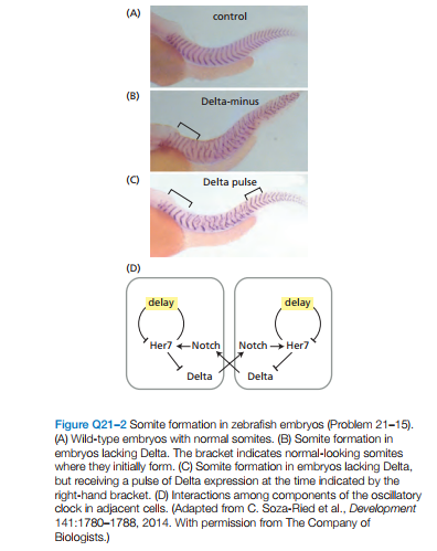
(A) control (B) Delta-minus (C) Delta pulse (D) delay delay Her7 +Notch Notch Her7 Delta Delta Figure Q21-2 Somite formation in zebrafish embryos (Problem 21-15). (A) Wild-type embryos with normal somites. (B) Somite formation in embryos lacking Delta. The bracket indicates normal-looking somites where they initially form. (C) Somite formation in embryos lacking Delta, but receiving a pulse of Delta expression at the time indicated by the right-hand bracket. (D) Interactions among components of the oscillatory clock in adjacent cells. (Adapted from C. Soza-Ried et al., Development 141:1780-1788, 2014. With permission from The Company of Biologists.)


The oscillatory clock that drives somite forma-
tion in vertebrates involves three essential components
Her7 (an unstable repressor of its own synthesis), Delta (a
transmembrane signaling molecule), and Notch (a trans-
membrane receptor for Delta). Notch is bound by Delta on
neighboring cells, activating the Notch signaling pathway,
which then activates Her7 transcription. Normally, this
system works flawlessly to create sharply defined somites
(Figure Q21–2A). In the absence of Delta, however, only
the first five somites form normally, and the rest are poorly
defined (Figure Q21–2B). If a pulse of Delta is supplied
later, somite formation returns to normal in the regions
where Delta was present (Figure Q21–2C). A diagram of
the connections between the components of the clock
and how they interact in adjacent cells is shown in Figure
Q21–2D. In the absence of Delta, why do the cells become
unsynchronized? What is it about the presence of Delta
that keeps adjacent cells oscillating in synchrony?

Step by step
Solved in 2 steps


