B. subtilis 1X3-4 micron • Notice: most cells appear purple while a few appear pink (arrow) Review fig 3.46 on page 171 to explain this phenomenon.
B. subtilis 1X3-4 micron • Notice: most cells appear purple while a few appear pink (arrow) Review fig 3.46 on page 171 to explain this phenomenon.

Biology: The Dynamic Science (MindTap Course List)
4th Edition
ISBN:9781305389892
Author:Peter J. Russell, Paul E. Hertz, Beverly McMillan
Publisher:Peter J. Russell, Paul E. Hertz, Beverly McMillan
Chapter49: Animal Reproduction
Section: Chapter Questions
Problem 14TYK

Related questions
Question
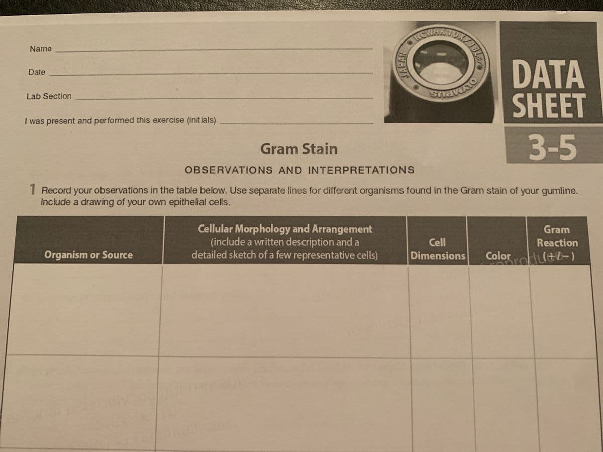
Transcribed Image Text:Name
DATA
SHEET
Date
Lab Section
I was present and performed this exercise (initials)
3-5
Gram Stain
OBSERVATIONS AND INTERPRETATIONS
1 Record your observations in the table below. Use separate lines for different organisms found in the Gram stain of your gumline.
Include a drawing of your own epithelial cells.
Cellular Morphology and Arrangement
(include a written description and a
detailed sketch of a few representative cells)
Gram
Cell
Reaction
Organism or Source
Dimensions
Color
BL
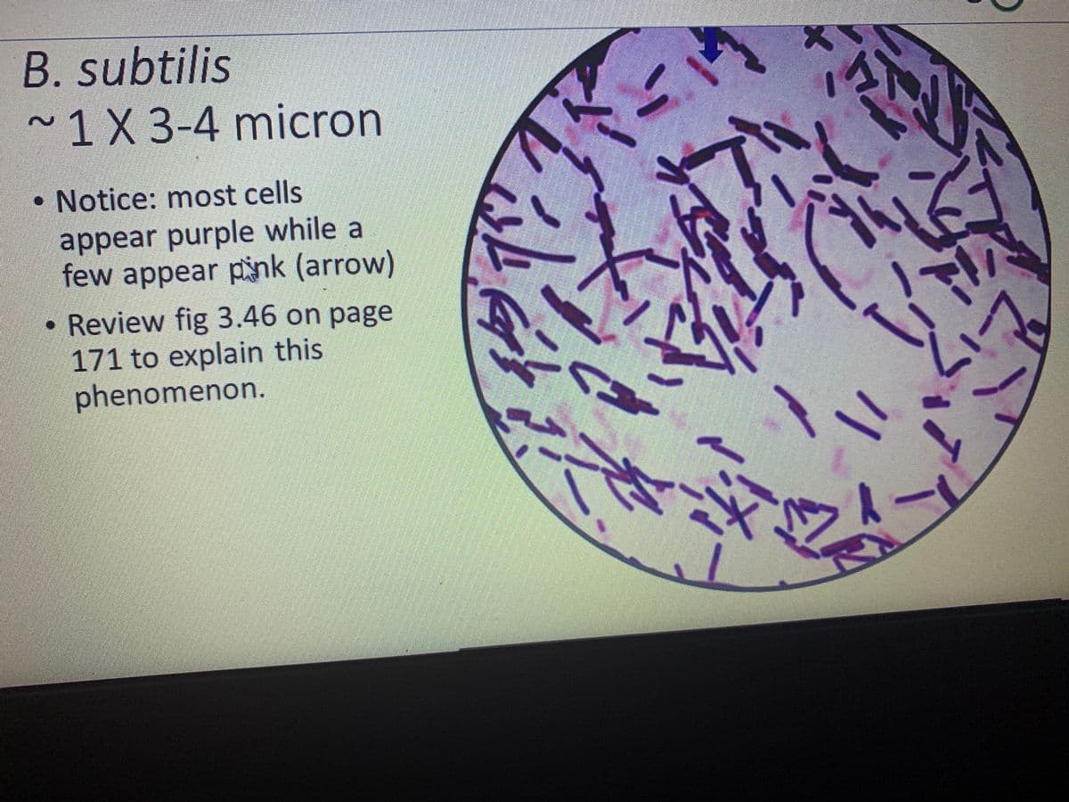
Transcribed Image Text:B. subtilis
~1X3-4 micron
• Notice: most cells
appear purple while a
few appear pink (arrow)
Review fig 3.46 on page
171 to explain this
phenomenon.
Expert Solution
Step 1
Gram staining is a common technique used to distinguish between two large groups of bacteria based on their different cell wall constituents. Gram staining distinguishes between Gram-positive and Gram-negative classes by painting these cells red or purple. Gram-positive bacteria stain violet due to the presence of a dense coating of peptidoglycan in their cell walls, which preserves the purple crystal with which these cells are stained. Alternatively, Gram-negative bacteria stain red, which is due to the thinner peptidoglycan wall, which does not maintain the purple crystal during the decoloration process.

Step by step
Solved in 2 steps with 1 images

Knowledge Booster


Learn more about
Need a deep-dive on the concept behind this application? Look no further. Learn more about this topic, biology and related others by exploring similar questions and additional content below.Recommended textbooks for you
Biology: The Dynamic Science (MindTap Course List)
Biology
ISBN:
9781305389892
Author:
Peter J. Russell, Paul E. Hertz, Beverly McMillan
Publisher:
Cengage Learning

Biology: The Unity and Diversity of Life (MindTap…
Biology
ISBN:
9781305073951
Author:
Cecie Starr, Ralph Taggart, Christine Evers, Lisa Starr
Publisher:
Cengage Learning

Biology Today and Tomorrow without Physiology (Mi…
Biology
ISBN:
9781305117396
Author:
Cecie Starr, Christine Evers, Lisa Starr
Publisher:
Cengage Learning
Biology: The Dynamic Science (MindTap Course List)
Biology
ISBN:
9781305389892
Author:
Peter J. Russell, Paul E. Hertz, Beverly McMillan
Publisher:
Cengage Learning
Biology: The Unity and Diversity of Life (MindTap…
Biology
ISBN:
9781305073951
Author:
Cecie Starr, Ralph Taggart, Christine Evers, Lisa Starr
Publisher:
Cengage Learning
Biology Today and Tomorrow without Physiology (Mi…
Biology
ISBN:
9781305117396
Author:
Cecie Starr, Christine Evers, Lisa Starr
Publisher:
Cengage Learning

Anatomy & Physiology
Biology
ISBN:
9781938168130
Author:
Kelly A. Young, James A. Wise, Peter DeSaix, Dean H. Kruse, Brandon Poe, Eddie Johnson, Jody E. Johnson, Oksana Korol, J. Gordon Betts, Mark Womble
Publisher:
OpenStax College

Biology 2e
Biology
ISBN:
9781947172517
Author:
Matthew Douglas, Jung Choi, Mary Ann Clark
Publisher:
OpenStax

Biology (MindTap Course List)
Biology
ISBN:
9781337392938
Author:
Eldra Solomon, Charles Martin, Diana W. Martin, Linda R. Berg
Publisher:
Cengage Learning