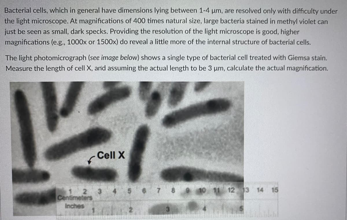
Bacterial cells, which in general have dimensions lying between 1-4 um, are resolved only with difficulty under the light microscope. At magnifications of 400 times natural size, large bacteria stained in methyl violet can just be seen as small, dark specks. Providing the resolution of the light microscope is good, higher magnifications (e.g., 1000x or 1500x) do reveal a little more of the internal structure of bacterial cells. The light photomicrograph (see image below) shows a single type of bacterial cell treated with Giemsa stain. Measure the length of cell X, and assuming the actual length to be 3 um, calculate the actual magnification. - Cell X 2 11 12 13 14 15 2 3 Centimeters Inches
Bacterial cells, which in general have dimensions lying between 1-4 um, are resolved only with difficulty under the light microscope. At magnifications of 400 times natural size, large bacteria stained in methyl violet can just be seen as small, dark specks. Providing the resolution of the light microscope is good, higher magnifications (e.g., 1000x or 1500x) do reveal a little more of the internal structure of bacterial cells. The light photomicrograph (see image below) shows a single type of bacterial cell treated with Giemsa stain. Measure the length of cell X, and assuming the actual length to be 3 um, calculate the actual magnification. - Cell X 2 11 12 13 14 15 2 3 Centimeters Inches

Chapter2: Aquatic Plants And Animals
Section: Chapter Questions
Problem 2KA

Related questions

Question
Answer these questions.
1. What is the actual magnification of Cell X?
2. How has magnification of this number of times been achieved if the light microscope itself (used in taking the photograph) only magnifies 1500 times?
Transcribed Image Text:Bacterial cells, which in general have dimensions lying between 1-4 um, are resolved only with difficulty under
the light microscope. At magnifications of 400 times natural size, large bacteria stained in methyl violet can
just be seen as small, dark specks. Providing the resolution of the light microscope is good, higher
magnifications (e.g., 1000x or 1500x) do reveal a little more of the internal structure of bacterial cells.
The light photomicrograph (see image below) shows a single type of bacterial cell treated with Giemsa stain.
Measure the length of cell X, and assuming the actual length to be 3 um, calculate the actual magnification.
- Cell X
2 11 12 13 14 15
2 3
Centimeters
Inches
Expert Solution
This question has been solved!
Explore an expertly crafted, step-by-step solution for a thorough understanding of key concepts.

Step by step
Solved in 2 steps

Knowledge Booster


Learn more about
Need a deep-dive on the concept behind this application? Look no further. Learn more about this topic, biology and related others by exploring similar questions and additional content below.Recommended textbooks for you