
Biology 2e
2nd Edition
ISBN:9781947172517
Author:Matthew Douglas, Jung Choi, Mary Ann Clark
Publisher:Matthew Douglas, Jung Choi, Mary Ann Clark
Chapter22: Prokaryotes: Bacteria And Archaea
Section: Chapter Questions
Problem 24RQ: A person in England arrives at a medical clinic with a fever and swollen lymph nodes shortly after...

Related questions

Question
Can S-layer proteins be detected by immunolabelling when a capsule is present? How do you know?
I need help finding the answer in the article and explain in short answer
based of the answer in the article
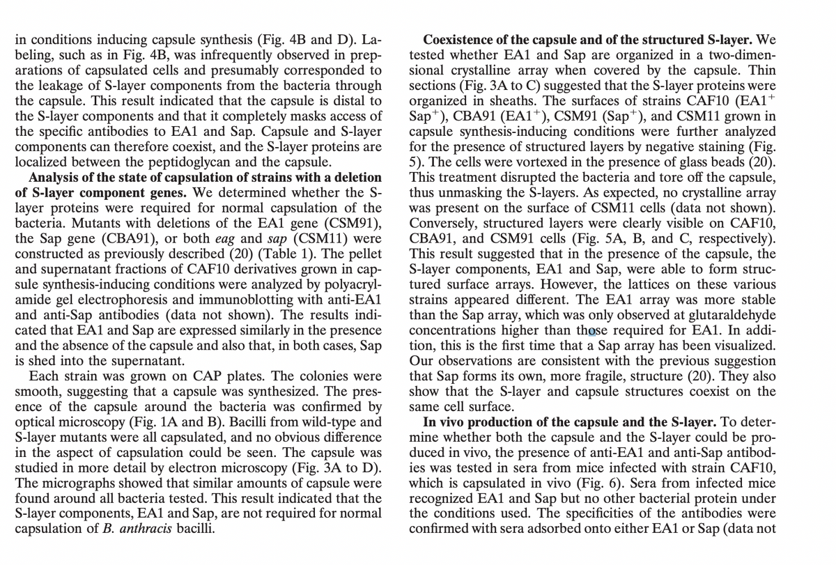
Transcribed Image Text:in conditions inducing capsule synthesis (Fig. 4B and D). La-
beling, such as in Fig. 4B, was infrequently observed in prep-
arations of capsulated cells and presumably corresponded to
the leakage of S-layer components from the bacteria through
the capsule. This result indicated that the capsule is distal to
the S-layer components and that it completely masks access of
the specific antibodies to EA1 and Sap. Capsule and S-layer
components can therefore coexist, and the S-layer proteins are
localized between the peptidoglycan and the capsule.
Analysis of the state of capsulation of strains with a deletion
of S-layer component genes. We determined whether the S-
layer proteins were required for normal capsulation of the
bacteria. Mutants with deletions of the EA1 gene (CSM91),
the Sap gene (CBA91), or both eag and sap (CSM11) were
constructed as previously described (20) (Table 1). The pellet
and supernatant fractions of CAF10 derivatives grown in cap-
sule synthesis-inducing conditions were analyzed by polyacryl-
amide gel electrophoresis and immunoblotting with anti-EA1
and anti-Sap antibodies (data not shown). The results indi-
cated that EA1 and Sap are expressed similarly in the presence
and the absence of the capsule and also that, in both cases, Sap
is shed into the supernatant.
Each strain was grown on CAP plates. The colonies were
smooth, suggesting that a capsule was synthesized. The pres-
ence of the capsule around the bacteria was confirmed by
optical microscopy (Fig. 1A and B). Bacilli from wild-type and
S-layer mutants were all capsulated, and no obvious difference
in the aspect of capsulation could be seen. The capsule was
studied in more detail by electron microscopy (Fig. 3A to D).
The micrographs showed that similar amounts of capsule were
found around all bacteria tested. This result indicated that the
S-layer components, EA1 and Sap, are not required for normal
capsulation of B. anthracis bacilli.
Coexistence of the capsule and of the structured S-layer. We
tested whether EA1 and Sap are organized in a two-dimen-
sional crystalline array when covered by the capsule. Thin
sections (Fig. 3A to C) suggested that the S-layer proteins were
organized in sheaths. The surfaces of strains CAF10 (EA1+
Sap*), CBA91 (EA1+), CSM91 (Sap*), and CSM11 grown in
capsule synthesis-inducing conditions were further analyzed
for the presence of structured layers by negative staining (Fig.
5). The cells were vortexed in the presence of glass beads (20).
This treatment disrupted the bacteria and tore off the capsule,
thus unmasking the S-layers. As expected, no crystalline array
was present on the surface of CSM11 cells (data not shown).
Conversely, structured layers were clearly visible on CAF10,
CBA91, and CSM91 cells (Fig. 5A, B, and C, respectively).
This result suggested that in the presence of the capsule, the
S-layer components, EA1 and Sap, were able to form struc-
tured surface arrays. However, the lattices on these various
strains appeared different. The EA1 array was more stable
than the Sap array, which was only observed at glutaraldehyde
concentrations higher than those required for EA1. In addi-
tion, this is the first time that a Sap array has been visualized.
Our observations are consistent with the previous suggestion
that Sap forms its own, more fragile, structure (20). They also
show that the S-layer and capsule structures coexist on the
same cell surface.
In vivo production of the capsule and the S-layer. To deter-
mine whether both the capsule and the S-layer could be pro-
duced in vivo, the presence of anti-EA1 and anti-Sap antibod-
ies was tested in sera from mice infected with strain CAF10,
which is capsulated in vivo (Fig. 6). Sera from infected mice
recognized EA1 and Sap but no other bacterial protein under
the conditions used. The specificities of the antibodies were
confirmed with sera adsorbed onto either EA1 or Sap (data not
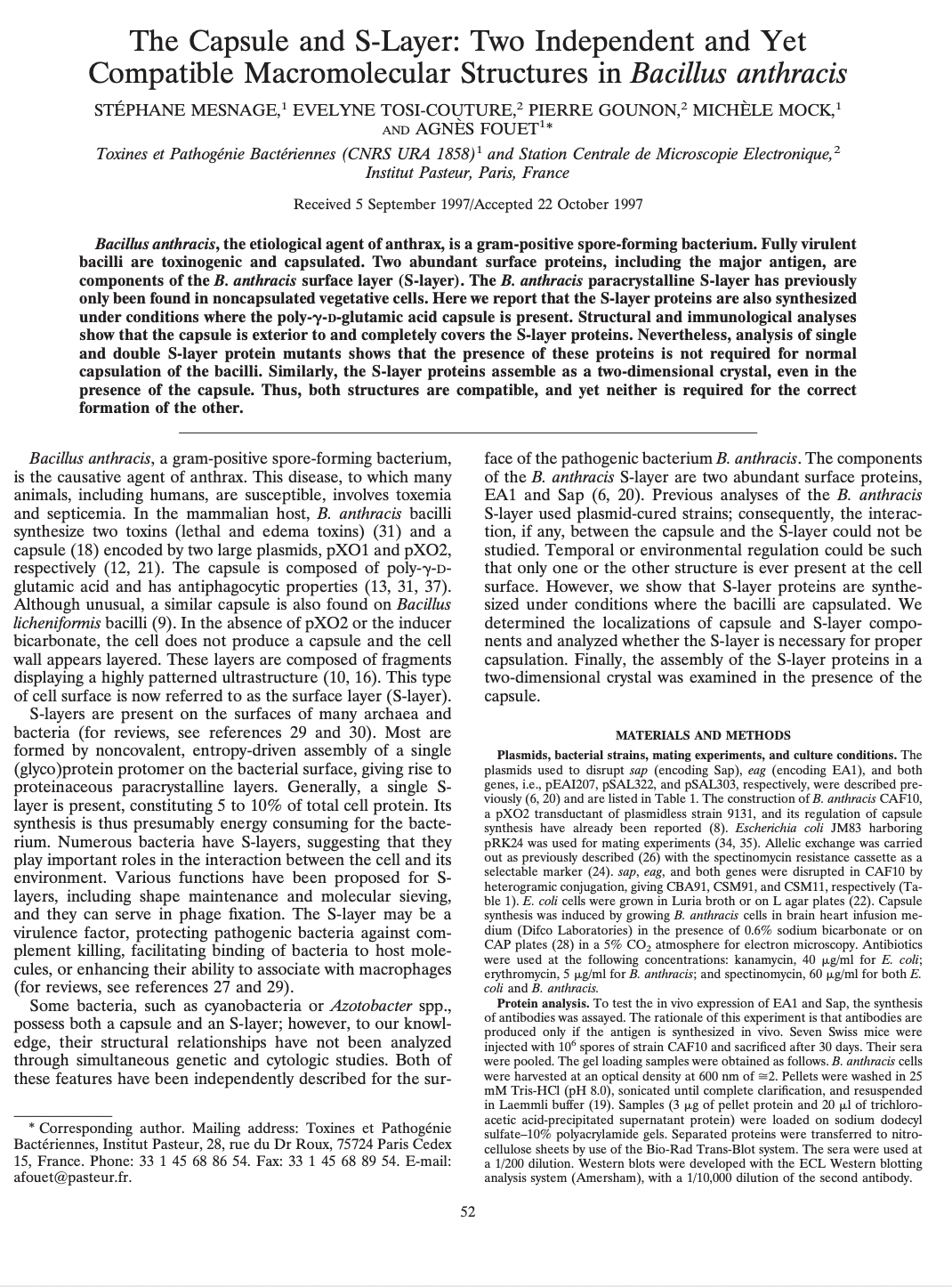
Transcribed Image Text:The Capsule and S-Layer: Two Independent and Yet
Compatible Macromolecular Structures in Bacillus anthracis
STÉPHANE MESNAGE,¹ EVELYNE TOSI-COUTURE,² PIERRE GOUNON,² MICHÈLE MOCK,¹
AND AGNES FOUET¹*
Toxines et Pathogénie Bactériennes (CNRS URA 1858)¹ and Station Centrale de Microscopie Electronique, ²
Institut Pasteur, Paris, France
Received 5 September 1997/Accepted 22 October 1997
Bacillus anthracis, the etiological agent of anthrax, is a gram-positive spore-forming bacterium. Fully virulent
bacilli are toxinogenic and capsulated. Two abundant surface proteins, including the major antigen, are
components of the B. anthracis surface layer (S-layer). The B. anthracis paracrystalline S-layer has previously
only been found in noncapsulated vegetative cells. Here we report that the S-layer proteins are also synthesized
under conditions where the poly-y-D-glutamic acid capsule is present. Structural and immunological analyses
show that the capsule is exterior to and completely covers the S-layer proteins. Nevertheless, analysis of single
and double S-layer protein mutants shows that the presence of these proteins is not required for normal
capsulation of the bacilli. Similarly, the S-layer proteins assemble as a two-dimensional crystal, even in the
presence of the capsule. Thus, both structures are compatible, and yet neither is required for the correct
formation of the other.
Bacillus anthracis, a gram-positive spore-forming bacterium,
is the causative agent of anthrax. This disease, to which many
animals, including humans, are susceptible, involves toxemia
and septicemia. In the mammalian host, B. anthracis bacilli
synthesize two toxins (lethal and edema toxins) (31) and a
capsule (18) encoded by two large plasmids, pX01 and pXO2,
respectively (12, 21). The capsule is composed of poly-y-D-
glutamic acid and has antiphagocytic properties (13, 31, 37).
Although unusual, a similar capsule is also found on Bacillus
licheniformis bacilli (9). In the absence of pXO2 or the inducer
bicarbonate, the cell does not produce a capsule and the cell
wall appears layered. These layers are composed of fragments
displaying a highly patterned ultrastructure (10, 16). This type
of cell surface is now referred to as the surface layer (S-layer).
S-layers are present on the surfaces of many archaea and
bacteria (for reviews, see references 29 and 30). Most are
formed by noncovalent, entropy-driven assembly of a single
(glyco)protein protomer on the bacterial surface, giving rise to
proteinaceous paracrystalline layers. Generally, a single S-
layer is present, constituting 5 to 10% of total cell protein. Its
synthesis is thus presumably energy consuming for the bacte-
rium. Numerous bacteria have S-layers, suggesting that they
play important roles in the interaction between the cell and its
environment. Various functions have been proposed for S-
layers, including shape maintenance and molecular sieving,
and they can serve in phage fixation. The S-layer may be a
virulence factor, protecting pathogenic bacteria against com-
plement killing, facilitating binding of bacteria to host mole-
cules, or enhancing their ability to associate with macrophages
(for reviews, see references 27 and 29).
Some bacteria, such as cyanobacteria or Azotobacter spp.,
possess both a capsule and an S-layer; however, to our knowl-
edge, their structural relationships have not been analyzed
through simultaneous genetic and cytologic studies. Both of
these features have been independently described for the sur-
* Corresponding author. Mailing address: Toxines et Pathogénie
Bactériennes, Institut Pasteur, 28, rue du Dr Roux, 75724 Paris Cedex
15, France. Phone: 33 1 45 68 86 54. Fax: 33 1 45 68 89 54. E-mail:
afouet@pasteur.fr.
52
face of the pathogenic bacterium B. anthracis. The components
of the B. anthracis S-layer are two abundant surface proteins,
EA1 and Sap (6, 20). Previous analyses of the B. anthracis
S-layer used plasmid-cured strains; consequently, the interac-
tion, if any, between the capsule and the S-layer could not be
studied. Temporal or environmental regulation could be such
that only one or the other structure is ever present at the cell
surface. However, we show that S-layer proteins are synthe-
sized under conditions where the bacilli are capsulated. We
determined the localizations of capsule and S-layer compo-
nents and analyzed whether the S-layer is necessary for proper
capsulation. Finally, the assembly of the S-layer proteins in a
two-dimensional crystal was examined in the presence of the
capsule.
MATERIALS AND METHODS
Plasmids, bacterial strains, mating experiments, and culture conditions. The
plasmids used to disrupt sap (encoding Sap), eag (encoding EA1), and both
genes, i.e., PEA1207, pSAL322, and pSAL303, respectively, were described pre-
viously (6, 20) and are listed in Table 1. The construction of B. anthracis CAF10,
a pXO2 transductant of plasmidless strain 9131, and its regulation of capsule
synthesis have already been reported (8). Escherichia coli JM83 harboring
pRK24 was used for mating experiments (34, 35). Allelic exchange was carried
out as previously described (26) with the spectinomycin resistance cassette as a
selectable marker (24). sap, eag, and both genes were disrupted in CAF10 by
heterogramic conjugation, giving CBA91, CSM91, and CSM11, respectively (Ta-
ble 1). E. coli cells were grown in Luria broth on agar plates (22). Capsule
synthesis was induced by growing B. anthracis cells in brain heart infusion me-
dium (Difco Laboratories) in the presence of 0.6% sodium bicarbonate or on
CAP plates (28) in a 5% CO₂ atmosphere for electron microscopy. Antibiotics
were used at the following concentrations: kanamycin, 40 µg/ml for E. coli;
erythromycin, 5 µg/ml for B. anthracis; and spectinomycin, 60 µg/ml for both E.
coli and B. anthracis.
you w
Protein analysis. To test the in vivo expression of EA1 and Sap, the synthesis
of antibodies was assayed. The rationale of this experiment is that antibodies are
produced only if the antigen is synthesized in vivo. Seven Swiss mice were
injected with 106 spores of strain CAF10 and sacrificed after 30 days. Their sera
were pooled. The gel loading samples were obtained as follows. B. anthracis cells
were harvested at an
an optical density at 600 nm of =2. Pellets were washed in 25
mM Tris-HCl (pH 8.0), sonicated until complete clarification, and resuspended
in Laemmli buffer (19). Samples (3 μg of
of pellet protein and 20 μl of trichloro-
acetic acid-precipitated supernatant protein) were loaded on sodium dodecyl
sulfate-10% polyacrylamide gels. Separated proteins were transferred to nitro-
cellulose sheets by use of the Bio-Rad Trans-Blot system. The sera were used at
1/200 dilution. Western blots were developed with the ECL Western blotting
analysis system (Amersham), with a 1/10,000 dilution of the second antibody.
Expert Solution
This question has been solved!
Explore an expertly crafted, step-by-step solution for a thorough understanding of key concepts.

Step by step
Solved in 2 steps

Knowledge Booster


Learn more about
Need a deep-dive on the concept behind this application? Look no further. Learn more about this topic, biology and related others by exploring similar questions and additional content below.Recommended textbooks for you
Biology 2e
Biology
ISBN:
9781947172517
Author:
Matthew Douglas, Jung Choi, Mary Ann Clark
Publisher:
OpenStax

Biology 2e
Biology
ISBN:
9781947172517
Author:
Matthew Douglas, Jung Choi, Mary Ann Clark
Publisher:
OpenStax