Modified Lateral and Lateral Oblique If evaluation of the entire length of the urinary tract is of concern in a male, the hind limbs may mask the membra- nous and penile urethra' in a true lateral position. There are two alternatives that could be considered the modified lateral and lateral oblique. Modified Lateral Positioning Place In: Right lateral recumbency. Head: Keep in a natural position, and hold appropriately with a sandbag over the neck. Be careful not to restrict breathing. Forelimbs: Pull cranially and sandbag. Place a small foam pad between the forelimbs to help eliminate rotation of the cranial abdomen. Hind Limbs: Pull the pelvic limbs cranially as far forward as possible without causing rotation of the body from the table. An appropriately sized foam pad placed between the femurs may help eliminate rotation of the pelvis. Place sandbags over the limbs. BOTH POSITIONS Measure: Over the ischium. Central Ray: Over the cranial wing of the ilium unless the rectum is of interest; in that case have the central beam over the pelvis. Borders: For a bladder, prostate, or caudal contrast study, include L4 and the caudal aspect of the rectum. TECHNICIAN NOTES Remember to collimate to the area of interest and include the markers. FIGURE 18-8 Positioning for a modified lateral view of a male for contrast studies. The hind limbs are pulled cranially as far as possible for evaluation of the membranous and penile urethra. Sternum: Elevate the ventral abdomen with wedged sponges so the sternum is at the same plane as the vertebrae. Have the central ray be perpendicular to both. Comments and Tips Ensure that the pelvic limbs are not superimposed over the caudal aspect of the os penis. . Expose immediately at the end phase of expiration. Lateral Oblique Positioning Place in: Right lateral recumbency. Head: Keep in a natural position and support appropriately with a sandbag over the neck. Forelimbs: Pull cranially and sandbag. Hind Limbs: Pull the dependent limb caudally and place a sandbag over the femur to keep in position. Raise the contralateral limb so that it is pulled dorsally and out of the field of view. A bungee cord or gauze tied around the tarsus and metatarsus and secured to the table, machine or sandbag will help keep the limb out of the field of view. B LO-20' FIGURE 18-9 A, Positioning for lateral oblique view of the canine abdomen. Ensure that the pelvic limbs are not superimposed over the caudal aspect of the os penis. B, Lateral oblique radiograph of the abdomen of a feline patient during an excretory urography using iodine.
Modified Lateral and Lateral Oblique If evaluation of the entire length of the urinary tract is of concern in a male, the hind limbs may mask the membra- nous and penile urethra' in a true lateral position. There are two alternatives that could be considered the modified lateral and lateral oblique. Modified Lateral Positioning Place In: Right lateral recumbency. Head: Keep in a natural position, and hold appropriately with a sandbag over the neck. Be careful not to restrict breathing. Forelimbs: Pull cranially and sandbag. Place a small foam pad between the forelimbs to help eliminate rotation of the cranial abdomen. Hind Limbs: Pull the pelvic limbs cranially as far forward as possible without causing rotation of the body from the table. An appropriately sized foam pad placed between the femurs may help eliminate rotation of the pelvis. Place sandbags over the limbs. BOTH POSITIONS Measure: Over the ischium. Central Ray: Over the cranial wing of the ilium unless the rectum is of interest; in that case have the central beam over the pelvis. Borders: For a bladder, prostate, or caudal contrast study, include L4 and the caudal aspect of the rectum. TECHNICIAN NOTES Remember to collimate to the area of interest and include the markers. FIGURE 18-8 Positioning for a modified lateral view of a male for contrast studies. The hind limbs are pulled cranially as far as possible for evaluation of the membranous and penile urethra. Sternum: Elevate the ventral abdomen with wedged sponges so the sternum is at the same plane as the vertebrae. Have the central ray be perpendicular to both. Comments and Tips Ensure that the pelvic limbs are not superimposed over the caudal aspect of the os penis. . Expose immediately at the end phase of expiration. Lateral Oblique Positioning Place in: Right lateral recumbency. Head: Keep in a natural position and support appropriately with a sandbag over the neck. Forelimbs: Pull cranially and sandbag. Hind Limbs: Pull the dependent limb caudally and place a sandbag over the femur to keep in position. Raise the contralateral limb so that it is pulled dorsally and out of the field of view. A bungee cord or gauze tied around the tarsus and metatarsus and secured to the table, machine or sandbag will help keep the limb out of the field of view. B LO-20' FIGURE 18-9 A, Positioning for lateral oblique view of the canine abdomen. Ensure that the pelvic limbs are not superimposed over the caudal aspect of the os penis. B, Lateral oblique radiograph of the abdomen of a feline patient during an excretory urography using iodine.

Chapter15: The Urinary System
Section: Chapter Questions
Problem F1CRE

Related questions
Question
read the article that is in the images and write the most important data
Transcribed Image Text:11:51 p. m. Mar 28 mar.
192
web.whatsapp.com
PART TWO Radiographic Positioning and Related Anatomy
Modified Lateral and Lateral Oblique
If evaluation of the entire length of the urinary tract is of
concern in a male, the hind limbs may mask the membra-
nous and penile urethra' in a true lateral position. There are
two alternatives that could be considered the modified
lateral and lateral oblique.
Modified Lateral
Positioning
Place In: Right lateral recumbency.
Head: Keep in a natural position, and hold appropriately
with a sandbag over the neck. Be careful not to restrict
breathing.
Forelimbs: Pull cranially and sandbag. Place a small foam
pad between the forelimbs to help eliminate rotation of
the cranial abdomen.
Hind Limbs: Pull the pelvic limbs cranially as far forward as
possible without causing rotation of the body from the
table. An appropriately sized foam pad placed between
the femurs may help eliminate rotation of the pelvis. Place
sandbags over the limbs.
BOTH POSITIONS Measure: Over the ischium.
Central Ray: Over the cranial wing of the ilium unless the
rectum is of interest; in that case have the central beam
over the pelvis.
Borders: For a bladder, prostate, or caudal contrast study,
include L4 and the caudal aspect of the rectum.
TECHNICIAN NOTES Remember to collimate to the
area of interest and include the markers.
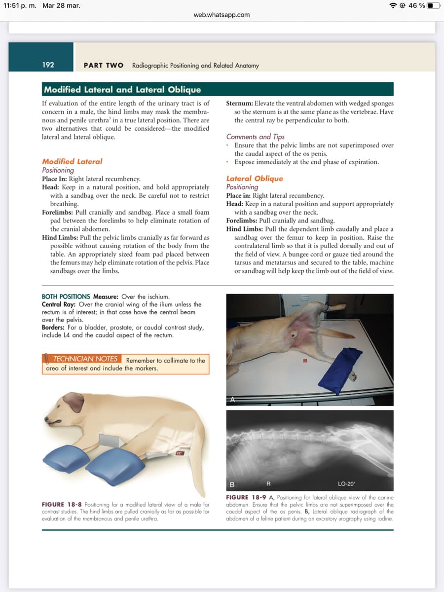
FIGURE 18-8 Positioning for a modified lateral view of a male for
contrast studies. The hind limbs are pulled cranially as far as possible for
evaluation of the membranous and penile urethra.
Sternum: Elevate the ventral abdomen with wedged sponges
so the sternum is at the same plane as the vertebrae. Have
the central ray be perpendicular to both.
Comments and Tips
• Ensure that the pelvic limbs are not superimposed over
the caudal aspect of the os penis.
• Expose immediately at the end phase of expiration.
Lateral Oblique
Positioning
Place in: Right lateral recumbency.
Head: Keep in a natural position and support appropriately
with a sandbag over the neck.
Forelimbs: Pull cranially and sandbag.
Hind Limbs: Pull the dependent limb caudally and place a
sandbag over the femur to keep in position. Raise the
contralateral limb so that it is pulled dorsally and out of
the field of view. A bungee cord or gauze tied around the
tarsus and metatarsus and secured to the table, machine
or sandbag will help keep the limb out of the field of view.
R
LO-20'
B
FIGURE 18-9 A, Positioning for lateral oblique view of the canine
abdomen. Ensure that the pelvic limbs are not superimposed over the
caudal aspect of the os penis. B, Lateral oblique radiograph of the
abdomen of a feline patient during an excretory urography using iodine.
46%
Transcribed Image Text:11:51 p. m. Mar 28 mar.
Modified Lateral and Lateral Oblique-cont'd
Comments and Tips
Ensure that the pelvic limbs are not superimposed over
the caudal aspect of the os penis.
Expose immediately at the end phase of expiration.
For male dogs, this view may be needed because on the
ventrodorsal view there may be superimposition of the
penis over the bladder.
web.whatsapp.com
• Oblique views can also be achieved by placing the patient
in the VD or DV positions and rotating the body 15-30
degrees. This moves the esophagus, stomach, colon and
urinary bladder away from the vertebrae to allow better
visualization.
194
The criteria for proper symmetry will not apply to the
oblique views.
R
CHAPTER 18 Small Animal Abdomen
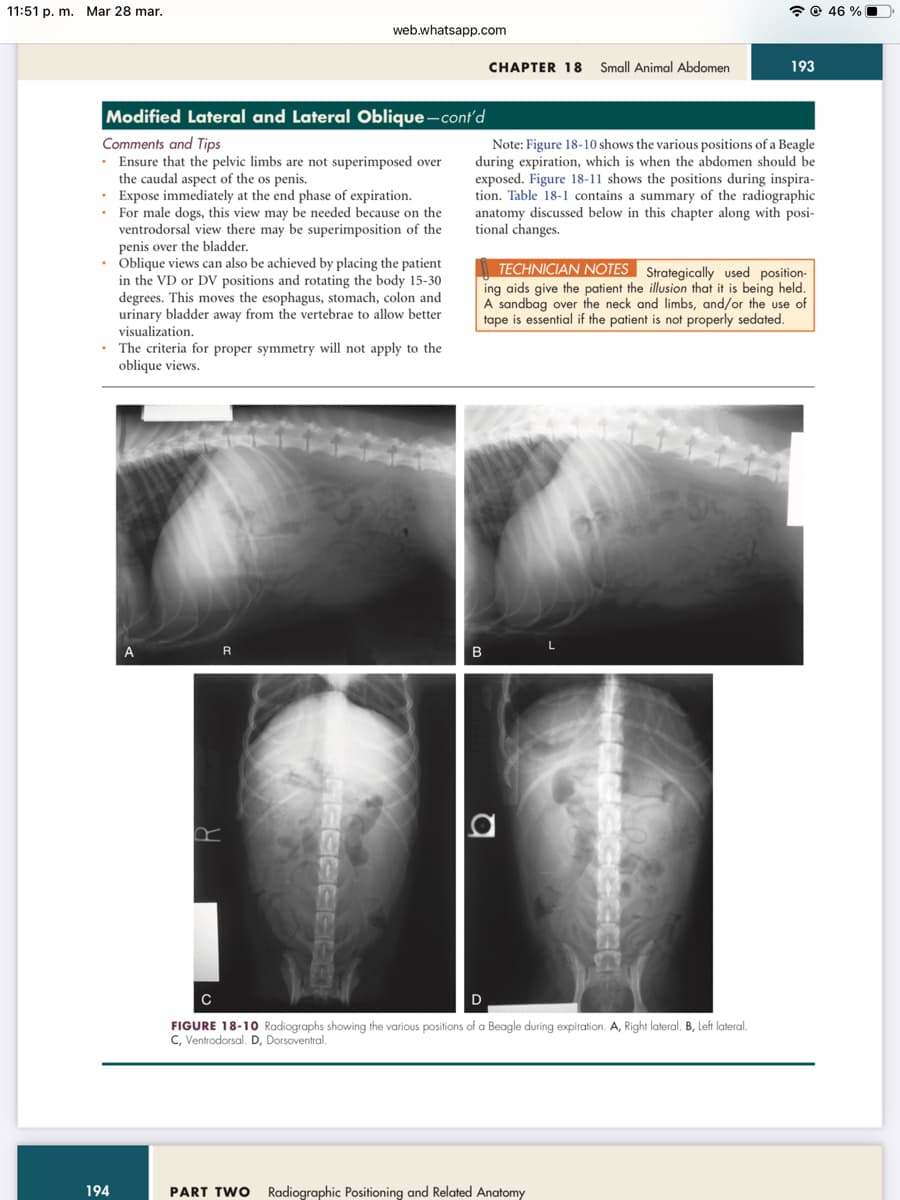
Note: Figure 18-10 shows the various positions of a Beagle
during expiration, which is when the abdomen should be
exposed. Figure 18-11 shows the positions during inspira-
tion. Table 18-1 contains a summary of the radiographic
anatomy discussed below in this chapter along with posi-
tional changes.
B
TECHNICIAN NOTES Strategically used position-
ing aids give the patient the illusion that it is being held.
A sandbag over the neck and limbs, and/or the use of
tape is essential if the patient is not properly sedated.
193
D
C
FIGURE 18-10 Radiographs showing the various positions of a Beagle during expiration. A, Right lateral. B, Left lateral.
C, Ventrodorsal. D, Dorsoventral.
PART TWO Radiographic Positioning and Related Anatomy
46%
Expert Solution
This question has been solved!
Explore an expertly crafted, step-by-step solution for a thorough understanding of key concepts.

This is a popular solution!

Trending now
This is a popular solution!

Step by step
Solved in 2 steps

Knowledge Booster


Learn more about
Need a deep-dive on the concept behind this application? Look no further. Learn more about this topic, biology and related others by exploring similar questions and additional content below.Recommended textbooks for you

Human Physiology: From Cells to Systems (MindTap …
Biology
ISBN:
9781285866932
Author:
Lauralee Sherwood
Publisher:
Cengage Learning
Human Physiology: From Cells to Systems (MindTap …
Biology
ISBN:
9781285866932
Author:
Lauralee Sherwood
Publisher:
Cengage Learning

Anatomy & Physiology
Biology
ISBN:
9781938168130
Author:
Kelly A. Young, James A. Wise, Peter DeSaix, Dean H. Kruse, Brandon Poe, Eddie Johnson, Jody E. Johnson, Oksana Korol, J. Gordon Betts, Mark Womble
Publisher:
OpenStax College