The cardiac output is low, but the left side shows hypertrophy. Explain how the cardiac output is usually compensated, and what has failed here.
The cardiac output is low, but the left side shows hypertrophy. Explain how the cardiac output is usually compensated, and what has failed here.

Biomedical Instrumentation Systems
1st Edition
ISBN:9781133478294
Author:Chatterjee
Publisher:Chatterjee
Chapter1: Introduction To Biomedical Instrumentation Systems
Section: Chapter Questions
Problem 1CS

Related questions
Question
The cardiac output is low, but the left side shows hypertrophy. Explain how the cardiac output
is usually compensated, and what has failed here.
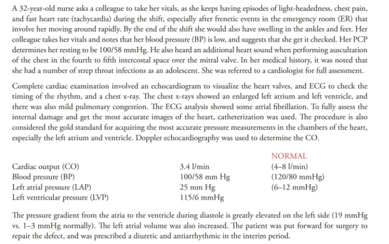
Transcribed Image Text:A 32-year-old nurse asks a colleague to take her vitals, as she keeps having episodes of light-headedness, chest pain,
and fast heart rate (tachycardia) during the shift, especially after frenetic events in the emergency room (ER) that
involve her moving around rapidly. By the end of the shift she would also have swelling in the ankles and feet. Her
colleague takes her vitals and notes that her blood pressure (BP) is low, and suggests that she get it checked. Her PCP
determines her resting to be 100/58 mmHg. He also heard an additional heart sound when performing auscultation
of the chest in the fourth to fifth intercostal space over the mitral valve. In her medical history, it was noted that
she had a number of strep throat infections as an adolescent. She was referred to a cardiologist for full assessment.
Complete cardiac examination involved an echocardiogram to visualize the heart valves, and ECG to check the
timing of the rhythm, and a chest x-ray. The chest x-rays showed an enlarged left atrium and left ventricle, and
there was also mild pulmonary congestion. The ECG analysis showed some atrial fibrillation. To fully assess the
internal damage and get the most accurate images of the heart, catheterization was used. The procedure is also
considered the gold standard for acquiring the most accurate pressure measurements in the chambers of the heart,
especially the left atrium and ventricle. Doppler echocardiography was used to determine the CO.
NORMAL
Cardiac output (CO)
Blood pressure (BP)
Left atrial pressure (LAP)
Left ventricular pressure (LVP)
3.4 l/min
100/58 mm Hg
25 mm Hg
115/6 mmHg
(4-8 l/min)
(120/80 mmHg)
(6–12 mmHg)
The pressure gradient from the atria to the ventricle during diastole is greatly elevated on the left side (19 mmHg
vs. 1-3 mmHg normally). The left atrial volume was also increased. The patient was put forward for surgery to
repair the defect, and was prescribed a diuretic and antiarrhythmic in the interim period.
Expert Solution
This question has been solved!
Explore an expertly crafted, step-by-step solution for a thorough understanding of key concepts.

Step by step
Solved in 6 steps

Recommended textbooks for you
Essentials of Pharmacology for Health Professions
Nursing
ISBN:
9781305441620
Author:
WOODROW
Publisher:
Cengage

Human Physiology: From Cells to Systems (MindTap …
Biology
ISBN:
9781285866932
Author:
Lauralee Sherwood
Publisher:
Cengage Learning
Essentials Health Info Management Principles/Prac…
Health & Nutrition
ISBN:
9780357191651
Author:
Bowie
Publisher:
Cengage