Through a series of experiments, we will now investigate how the human eye detects light in two conditions (in the presence of light and in the dark), in order to understand the patient impaired vision at night. The eye's retina is constituted by two types of photoreceptor cells: the rods and the cones. These cells can detect light and produce a cellular response through a specific signaling pathway. The internal structure of these photoreceptor cells is represented in the figure below. Rods differ from cones in that they are much taller, much thinner and have many membranous discs that play a key role in photoreception. Light is received by a membrane receptor, which initiates a signal transduction inside the cell. The cellular response produced consists of the release a neurotransmitter (glutamate) into a synaptic cleft, triggering a series of action potentials in the post-synaptic neuron (not shown in the figure). This electrical signal is then sent to (and processed by) the central nervous system to form an image in the brain.
Through a series of experiments, we will now investigate how the human eye detects light in two conditions (in the presence of light and in the dark), in order to understand the patient impaired vision at night. The eye's retina is constituted by two types of photoreceptor cells: the rods and the cones. These cells can detect light and produce a cellular response through a specific signaling pathway. The internal structure of these photoreceptor cells is represented in the figure below. Rods differ from cones in that they are much taller, much thinner and have many membranous discs that play a key role in photoreception. Light is received by a membrane receptor, which initiates a signal transduction inside the cell. The cellular response produced consists of the release a neurotransmitter (glutamate) into a synaptic cleft, triggering a series of action potentials in the post-synaptic neuron (not shown in the figure). This electrical signal is then sent to (and processed by) the central nervous system to form an image in the brain.

Biology: The Dynamic Science (MindTap Course List)
4th Edition
ISBN:9781305389892
Author:Peter J. Russell, Paul E. Hertz, Beverly McMillan
Publisher:Peter J. Russell, Paul E. Hertz, Beverly McMillan
Chapter39: Information Flow And The Neuron
Section: Chapter Questions
Problem 1TYK

Related questions

Question
Transcribed Image Text:Through a series of experiments, we will now investigate how the human eye detects light in two
conditions (in the presence of light and in the dark), in order to understand the patient impaired vision
at night.
The eye's retina is constituted by two types of photoreceptor cells: the rods and the cones. These cells
can detect light and produce a cellular response through a specific signaling pathway. The internal
structure of these photoreceptor cells is represented in the figure below. Rods differ from cones in that
they are much taller, much thinner and have many membranous discs that play a key role in
photoreception. Light is received by a membrane receptor, which initiates a signal transduction inside
the cell. The cellular response produced consists of the release a neurotransmitter (glutamate) into a
synaptic cleft, triggering a series of action potentials the post-synaptic neuron (not shown in the
figure). This electrical signal is then sent to (and processed by) the central nervous system to form an
image in the brain.
In rods, the signaling pathway involves a light-sensitive membrane receptor: a protein called
rhodopsin, which is responsible for detecting low light intensities (but not color).
In cones, the signaling pathway involves a color-sensitive membrane receptor: a protein called
photopsin, which is responsible for detecting light of different wavelengths (but is not able to
detect low light intensities).
Eye
GFP
Rod
(photoreceptor)
Path of light
Rhodopsin
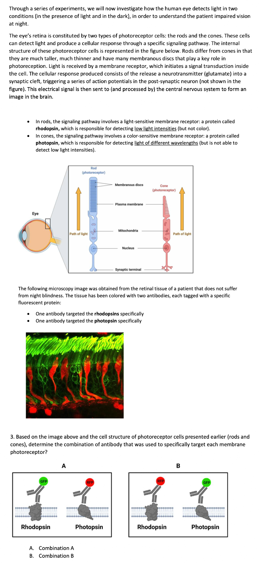
A
A. Combination A
B. Combination B
Membranous discs
One antibody targeted the rhodopsins specifically
One antibody targeted the photopsin specifically
Plasma membrane
The following microscopy image was obtained from the retinal tissue of a patient that does not suffer
from night blindness. The tissue has been colored with two antibodies, each tagged with a specific
fluorescent protein:
RFP
Mitochondria
Photopsin
Nucleus
3. Based on the image above and the cell structure of photoreceptor cells presented earlier (rods and
cones), determine the combination of antibody that was used to specifically target each membrane
photoreceptor?
Synaptic terminal
Cone
(photoreceptor)
Path of light
Rhodopsin
B
GFP
Photopsin
Expert Solution
This question has been solved!
Explore an expertly crafted, step-by-step solution for a thorough understanding of key concepts.

Step by step
Solved in 3 steps

Knowledge Booster


Learn more about
Need a deep-dive on the concept behind this application? Look no further. Learn more about this topic, biology and related others by exploring similar questions and additional content below.Recommended textbooks for you
Biology: The Dynamic Science (MindTap Course List)
Biology
ISBN:
9781305389892
Author:
Peter J. Russell, Paul E. Hertz, Beverly McMillan
Publisher:
Cengage Learning
Biology: The Dynamic Science (MindTap Course List)
Biology
ISBN:
9781305389892
Author:
Peter J. Russell, Paul E. Hertz, Beverly McMillan
Publisher:
Cengage Learning