VSV-GFP- SARS-CoV VSV-GFP- SARS-CoV-2 U Plaque Day 3 (duplicate) 1.51 1.0- 0.5- 0.0 VSV-GFP- SARS-CoV VSV-GFP- SARS-CoV-2 VSV-GFP- VSV-GFP- SARS-CoV SARS-CoV-2 Fig. 2. rVSV expressing SARS-CoV-2 spike spreads faster than does rVSV bearing SARS-CoV spike. Vero-E6 cells were infected with rVSV-GFP-SARS-CoV-2 or SARS-CoV (MOI = 0.01); 1 h postinfection, cells were washed with PBS and cultured in the presence of 1% methylcellulose. Photos were taken at 18 h and 24 h (A). After 72 h of infection, cells were fixed with 3.7% paraformaldehyde (PFA) and stained with crystal violet (B). The number and size of pla- ques are plotted in C and D, respectively. ***P<0.001. ns, not significant. Plaque number 80 60 40 20 18 h (10X) ns 24 h (20X) B D Plaque size (mm)
VSV-GFP- SARS-CoV VSV-GFP- SARS-CoV-2 U Plaque Day 3 (duplicate) 1.51 1.0- 0.5- 0.0 VSV-GFP- SARS-CoV VSV-GFP- SARS-CoV-2 VSV-GFP- VSV-GFP- SARS-CoV SARS-CoV-2 Fig. 2. rVSV expressing SARS-CoV-2 spike spreads faster than does rVSV bearing SARS-CoV spike. Vero-E6 cells were infected with rVSV-GFP-SARS-CoV-2 or SARS-CoV (MOI = 0.01); 1 h postinfection, cells were washed with PBS and cultured in the presence of 1% methylcellulose. Photos were taken at 18 h and 24 h (A). After 72 h of infection, cells were fixed with 3.7% paraformaldehyde (PFA) and stained with crystal violet (B). The number and size of pla- ques are plotted in C and D, respectively. ***P<0.001. ns, not significant. Plaque number 80 60 40 20 18 h (10X) ns 24 h (20X) B D Plaque size (mm)

Basic Clinical Laboratory Techniques 6E
6th Edition
ISBN:9781133893943
Author:ESTRIDGE
Publisher:ESTRIDGE
Chapter2: Basic Hematology
Section2.10: Principles Of Automated Hematology
Problem 2CS

Related questions

Question
Please help summarize
Transcribed Image Text:A
VSV-GFP-
SARS-CoV
VSV-GFP-
SARS-CoV-2
C
Plaque number
80
60-
40-
20
4¹.
0
18 h (10X)
ns
Plaque Day 3 (duplicate)
0.5-
0.0
VSV-GFP-
VSV-GFP-
SARS-CoV
VSV-GFP-
SARS-CoV-2
VSV-GFP-
SARS-CoV SARS-CoV-2
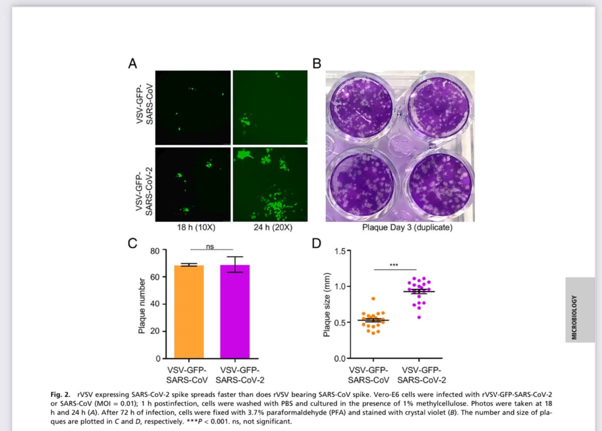
Fig. 2. rVSV expressing SARS-CoV-2 spike spreads faster than does rVSV bearing SARS-CoV spike. Vero-E6 cells were infected with rVSV-GFP-SARS-CoV-2
or SARS-CoV (MOI = 0.01); 1 h postinfection, cells were washed with PBS and cultured in the presence of 1% methylcellulose. Photos were taken at 18
h and 24 h (A). After 72 h of infection, cells were fixed with 3.7% paraformaldehyde (PFA) and stained with crystal violet (B). The number and size of pla-
ques are plotted in C and D, respectively. ***P<0.001. ns, not significant.
:2 "
24 h (20X)
B
D
Plaque size (mm)
1.51
1.0-
MICROBIOLOGY
Expert Solution
This question has been solved!
Explore an expertly crafted, step-by-step solution for a thorough understanding of key concepts.

Step by step
Solved in 2 steps

Knowledge Booster


Learn more about
Need a deep-dive on the concept behind this application? Look no further. Learn more about this topic, biology and related others by exploring similar questions and additional content below.Recommended textbooks for you