you are looking at a slide in the laboratory and observe a cell that occupies one quarter of the field of view at high magnification. use your field diameter calculation from lab activity 4 to estimate the size of this cell.
you are looking at a slide in the laboratory and observe a cell that occupies one quarter of the field of view at high magnification. use your field diameter calculation from lab activity 4 to estimate the size of this cell.

Chapter20: Radiology And Diagnostic Imaging
Section: Chapter Questions
Problem C1CRE

Related questions
Question
you are looking at a slide in the laboratory and observe a cell that occupies one quarter of the field of view at high magnification. use your field diameter calculation from lab activity 4 to estimate the size of this cell.
Transcribed Image Text:EXERCISE 4 Use of the Microscope
2. Use the coarse adjustment knob to bring the threads into
focus. Find the area where the threads overlap.
42
3. Rotate the nosepiece to select the low-power objective lens.
4. Use the fine adjustment knob to focus through the
overlapping threads. After determining which thread is on
top, which is in the middle, and which is on the bottom,
write your observations in the space provided.
Color of top thread
Color of middle thread
Color of bottom thread.
4
Relationship Between Magnification
and Field Diameter
At scanning magnification, the diameter of the field of view is
large and most of the slide specimen is visible. As magnifica-
tion increases, the field diameter decreases, because at higher
power the objective lens is closer to the slide and magnifies a
smaller area. Figure 4.5 reviews the relationship between mag-
nification and field diameter.
Field diameter at scanning and low magnifications can
be measured using millimeter graph paper glued to a micro-
scope slide. By aligning a vertical marking on the paper with
the edge of the field and then counting the number of mil-
limeter (mm) squares across the field, you can determine the
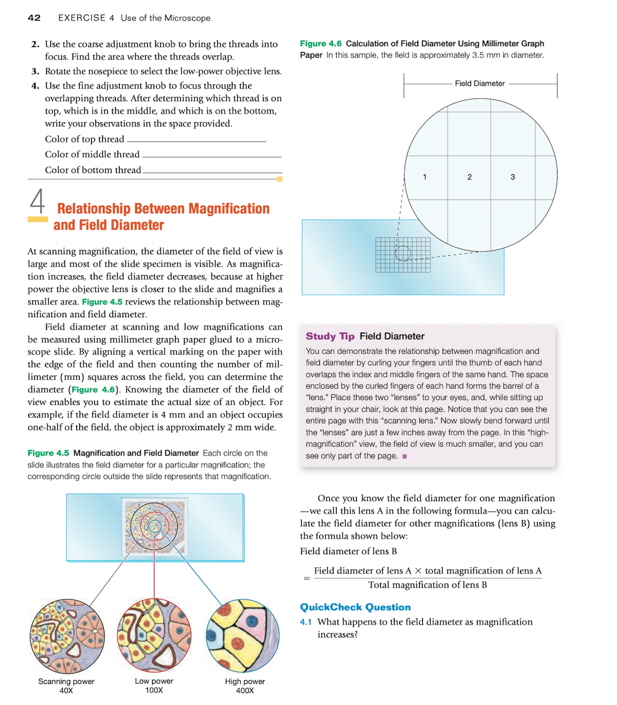
diameter (Figure 4.6). Knowing the diameter of the field of
view enables you to estimate the actual size of an object. For
example, if the field diameter is 4 mm and an object occupies
one-half of the field, the object is approximately 2 mm wide.
Figure 4.5 Magnification and Field Diameter Each circle on the
slide illustrates the field diameter for a particular magnification; the
corresponding circle outside the slide represents that magnification.
Scanning power
40X
Low power
100X
High power
400X
Figure 4.6 Calculation of Field Diameter Using Millimeter Graph
Paper In this sample, the field is approximately 3.5 mm in diameter.
1
Field Diameter
2
3
Study Tip Field Diameter
You can demonstrate the relationship between magnification and
field diameter by curling your fingers until the thumb of each hand
overlaps the index and middle fingers of the same hand. The space
enclosed by the curled fingers of each hand forms the barrel of a
"lens." Place these two "lenses" to your eyes, and, while sitting up
straight in your chair, look at this page. Notice that you can see the
entire page with this "scanning lens." Now slowly bend forward until
the "lenses" are just a few inches way from the page. In this "high-
magnification" view, the field of view is much smaller, and you can
see only part of the page.
Once you know the field diameter for one magnification
-we call this lens A in the following formula-you can calcu-
late the field diameter for other magnifications (lens B) using
the formula shown below:
Field diameter of lens B
Field diameter of lens A X total magnification of lens A
Total magnification of lens B
QuickCheck Question
4.1 What happens to the field diameter as magnification
increases?
Expert Solution
This question has been solved!
Explore an expertly crafted, step-by-step solution for a thorough understanding of key concepts.

This is a popular solution!

Trending now
This is a popular solution!

Step by step
Solved in 3 steps with 1 images

Knowledge Booster


Learn more about
Need a deep-dive on the concept behind this application? Look no further. Learn more about this topic, biology and related others by exploring similar questions and additional content below.Recommended textbooks for you
Understanding Health Insurance: A Guide to Billin…
Health & Nutrition
ISBN:
9781337679480
Author:
GREEN
Publisher:
Cengage
Understanding Health Insurance: A Guide to Billin…
Health & Nutrition
ISBN:
9781337679480
Author:
GREEN
Publisher:
Cengage