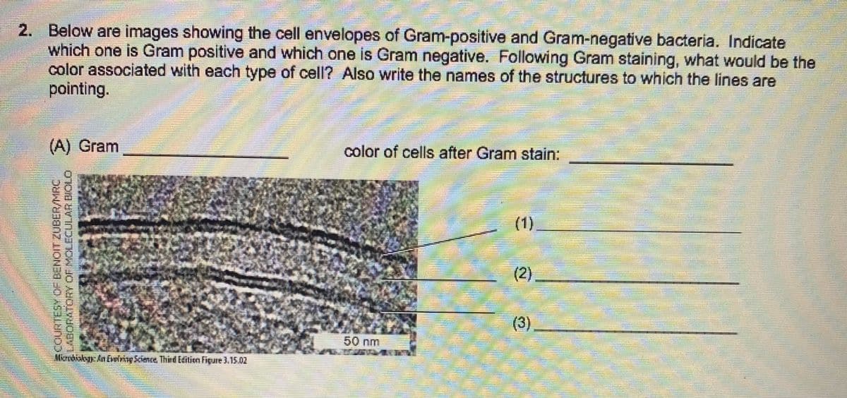
2. Below are images showing the cell envelopes of Gram-positive and Gram-negative bacteria. Indicate which one is Gram positive and which one is Gram negative. Following Gram staining, what would be the color associated with each type of cell? Also write the names of the structures to which the lines are pointing.
2. Below are images showing the cell envelopes of Gram-positive and Gram-negative bacteria. Indicate which one is Gram positive and which one is Gram negative. Following Gram staining, what would be the color associated with each type of cell? Also write the names of the structures to which the lines are pointing.

Concepts of Biology
1st Edition
ISBN:9781938168116
Author:Samantha Fowler, Rebecca Roush, James Wise
Publisher:Samantha Fowler, Rebecca Roush, James Wise
Chapter13: Diversity Of Microbes, Fungi, And Protists
Section: Chapter Questions
Problem 1ACQ: Figure 13.6 Which of the following statements is true? a. Gram-positive bacteria have a single cell...

Related questions
Question
Transcribed Image Text:2. Below are images showing the cell envelopes of Gram-positive and Gram-negative bacteria. Indicate
which one is Gram positive and which one is Gram negative. Following Gram staining, what would be the
color associated with each type of cell? Also write the names of the structures to which the lines are
pointing.
(A) Gram
color of cells after Gram stain:
(1)
(2).
(3)
50 nm
Moobiskyy AnEverimy Kience Third EHilien Figure 3.15.02
SCOURTESY OF BENOIT ZUBER/MRC.
O ORYOF MOLECULAR BIOLO
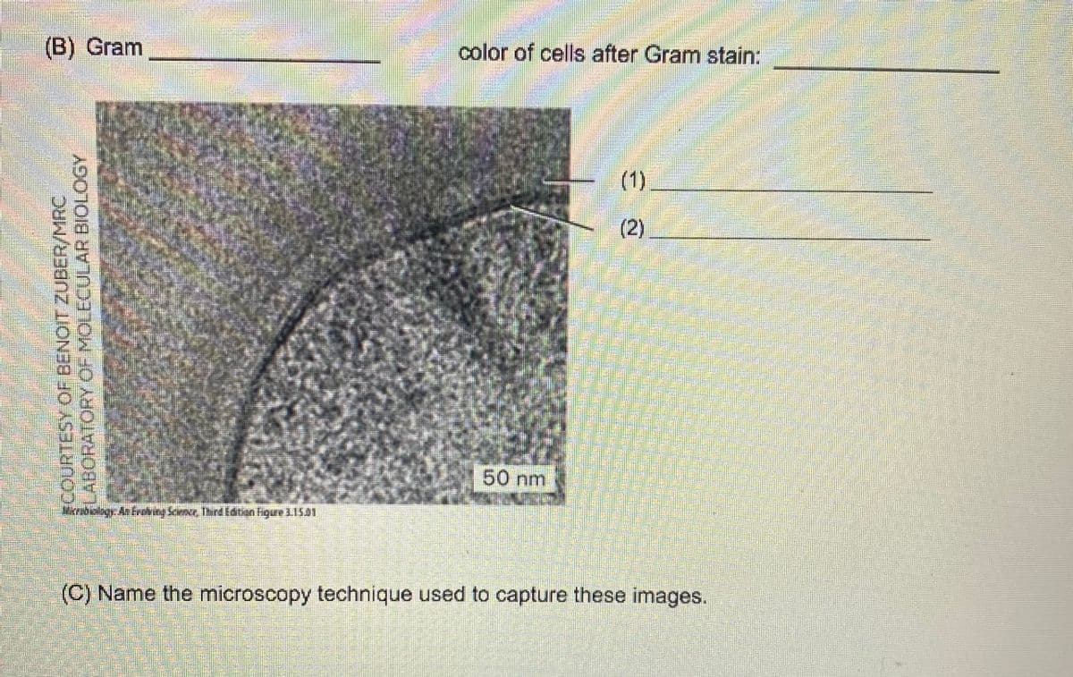
Transcribed Image Text:(B) Gram
color of cells after Gram stain:
(1)
(2)
50 nm
Merndiolagy An Ereving Science Thrd Edtion higure 31501
(C) Name the microscopy technique used to capture these images.
gcOURTESY OF BENOIT ZUDER/MRC
LABORATORY OF MOLECULAR BIOLOGY
Expert Solution
This question has been solved!
Explore an expertly crafted, step-by-step solution for a thorough understanding of key concepts.

This is a popular solution!

Trending now
This is a popular solution!

Step by step
Solved in 3 steps with 2 images

Recommended textbooks for you
Concepts of Biology
Biology
ISBN:
9781938168116
Author:
Samantha Fowler, Rebecca Roush, James Wise
Publisher:
OpenStax College

Biology 2e
Biology
ISBN:
9781947172517
Author:
Matthew Douglas, Jung Choi, Mary Ann Clark
Publisher:
OpenStax

Concepts of Biology
Biology
ISBN:
9781938168116
Author:
Samantha Fowler, Rebecca Roush, James Wise
Publisher:
OpenStax College
Biology 2e
Biology
ISBN:
9781947172517
Author:
Matthew Douglas, Jung Choi, Mary Ann Clark
Publisher:
OpenStax