Cerebral Hemispheres and Ventricles. 1. The outer gray matter of the cerebrum is called the divided into four lobes. The four major lobes of the cortex are the and is and lobes. 2. The parietal and frontal lobes are separated by the sulcus. 3. The sulcus separates the temporal lobe from the parietal and frontal lobes. 4. The motor cortex is located in the gyrus of the lobe. 5. The cerebral hemispheres are separated by the fissure. 6. The elevated ridges along the surface of the cerebral hemispheres are called 7. The somatosensory cortex is located in the gyrus of the lobe. 8. The visual cortex is located in the lobe. 9. The auditory cortex is located in the lobe.
Cerebral Hemispheres and Ventricles. 1. The outer gray matter of the cerebrum is called the divided into four lobes. The four major lobes of the cortex are the and is and lobes. 2. The parietal and frontal lobes are separated by the sulcus. 3. The sulcus separates the temporal lobe from the parietal and frontal lobes. 4. The motor cortex is located in the gyrus of the lobe. 5. The cerebral hemispheres are separated by the fissure. 6. The elevated ridges along the surface of the cerebral hemispheres are called 7. The somatosensory cortex is located in the gyrus of the lobe. 8. The visual cortex is located in the lobe. 9. The auditory cortex is located in the lobe.

Anatomy & Physiology
1st Edition
ISBN:9781938168130
Author:Kelly A. Young, James A. Wise, Peter DeSaix, Dean H. Kruse, Brandon Poe, Eddie Johnson, Jody E. Johnson, Oksana Korol, J. Gordon Betts, Mark Womble
Publisher:Kelly A. Young, James A. Wise, Peter DeSaix, Dean H. Kruse, Brandon Poe, Eddie Johnson, Jody E. Johnson, Oksana Korol, J. Gordon Betts, Mark Womble
Chapter13: Anatomy Of The Nervous System
Section: Chapter Questions
Problem 2ILQ: Watch this video (http://openstaxcollege.org/l/whitematter) to learn about the white matter in the...

Related questions
Question
Transcribed Image Text:Cerebral Hemispheres and Ventricles.
1. The outer gray matter of the cerebrum is called the
divided into four lobes. The four major lobes of the cortex are the
and is
and
lobes.
2. The parietal and frontal lobes are separated by the
sulcus.
3. The
sulcus separates the temporal lobe from the parietal and frontal lobes.
4. The motor cortex is located in the
gyrus of the
lobe.
5. The cerebral hemispheres are separated by the
fissure.
6. The elevated ridges along the surface of the cerebral hemispheres are called
7. The somatosensory cortex is located in the
gyrus of the
lobe.
8. The visual cortex is located in the
lobe.
9.
The auditory cortex is located in the
lobe.
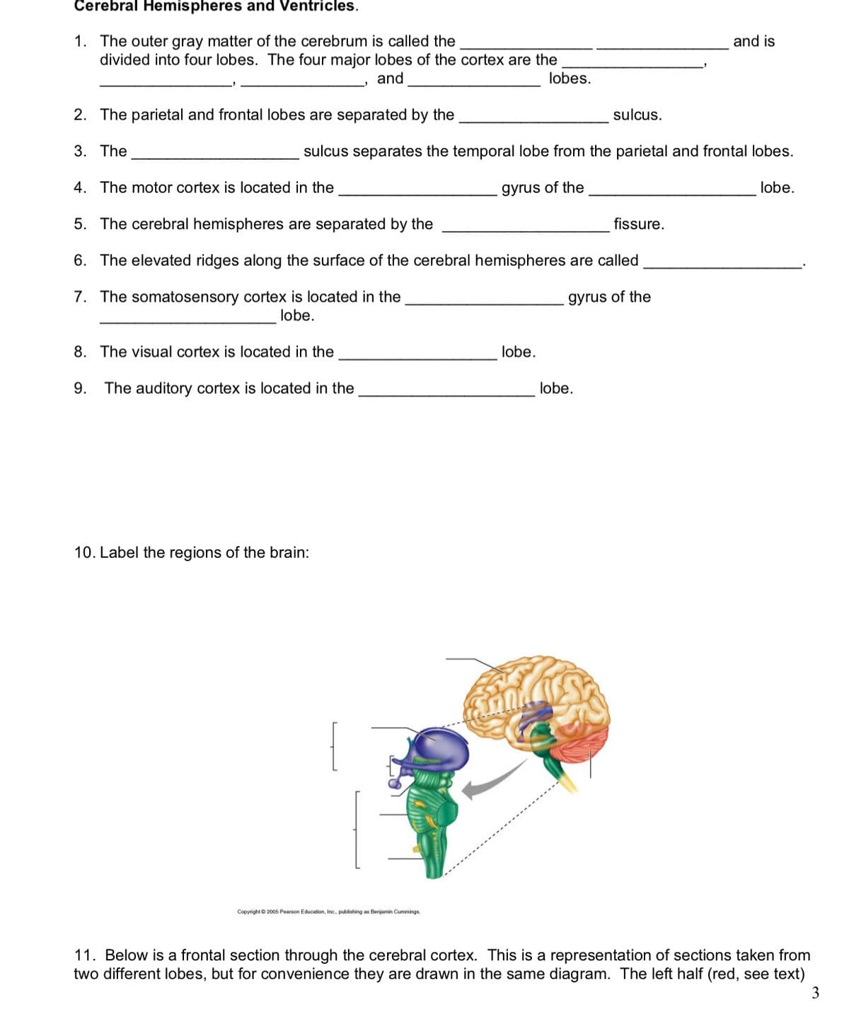
10. Label the regions of the brain:
Copyright © 2005 Pearson Education, Inc., publishing as Benjamin Cummings.
11. Below is a frontal section through the cerebral cortex. This is a representation of sections taken from
two different lobes, but for convenience they are drawn in the same diagram. The left half (red, see text)
3
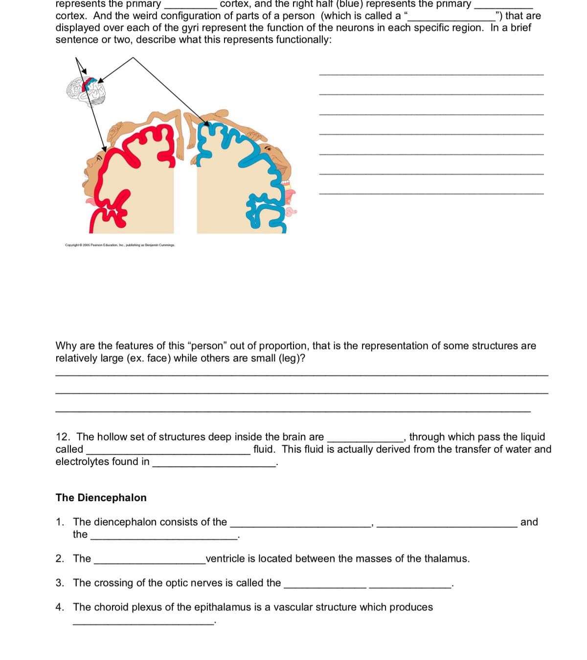
Transcribed Image Text:represents the primary
cortex. And the weird configuration of parts of a person (which is called a
displayed over each of the gyri represent the function of the neurons in each specific region. In a brief
sentence or two, describe what this represents functionally:
cortex, and the right half (blue) represents the primary
") that are
Copyright © 2005 Pearson Education, Inc., publishing as Benjamin Cummings.
Why are the features of this “person" out of proportion, that is the representation of some structures are
relatively large (ex. face) while others are small (leg)?
12. The hollow set of structures deep inside the brain are
called
through which pass the liquid
fluid. This fluid is actually derived from the transfer of water and
electrolytes found in
The Diencephalon
1. The diencephalon consists of the
the
and
2. The
ventricle is located between the masses of the thalamus.
3. The crossing of the optic nerves is called the
4. The choroid plexus of the epithalamus is a vascular structure which produces
Expert Solution
This question has been solved!
Explore an expertly crafted, step-by-step solution for a thorough understanding of key concepts.

This is a popular solution!

Trending now
This is a popular solution!

Step by step
Solved in 2 steps with 1 images

Recommended textbooks for you
Anatomy & Physiology
Biology
ISBN:
9781938168130
Author:
Kelly A. Young, James A. Wise, Peter DeSaix, Dean H. Kruse, Brandon Poe, Eddie Johnson, Jody E. Johnson, Oksana Korol, J. Gordon Betts, Mark Womble
Publisher:
OpenStax College

Human Physiology: From Cells to Systems (MindTap …
Biology
ISBN:
9781285866932
Author:
Lauralee Sherwood
Publisher:
Cengage Learning

Biology 2e
Biology
ISBN:
9781947172517
Author:
Matthew Douglas, Jung Choi, Mary Ann Clark
Publisher:
OpenStax
Anatomy & Physiology
Biology
ISBN:
9781938168130
Author:
Kelly A. Young, James A. Wise, Peter DeSaix, Dean H. Kruse, Brandon Poe, Eddie Johnson, Jody E. Johnson, Oksana Korol, J. Gordon Betts, Mark Womble
Publisher:
OpenStax College
Human Physiology: From Cells to Systems (MindTap …
Biology
ISBN:
9781285866932
Author:
Lauralee Sherwood
Publisher:
Cengage Learning
Biology 2e
Biology
ISBN:
9781947172517
Author:
Matthew Douglas, Jung Choi, Mary Ann Clark
Publisher:
OpenStax

Fundamentals of Sectional Anatomy: An Imaging App…
Biology
ISBN:
9781133960867
Author:
Denise L. Lazo
Publisher:
Cengage Learning

Biology: The Dynamic Science (MindTap Course List)
Biology
ISBN:
9781305389892
Author:
Peter J. Russell, Paul E. Hertz, Beverly McMillan
Publisher:
Cengage Learning
