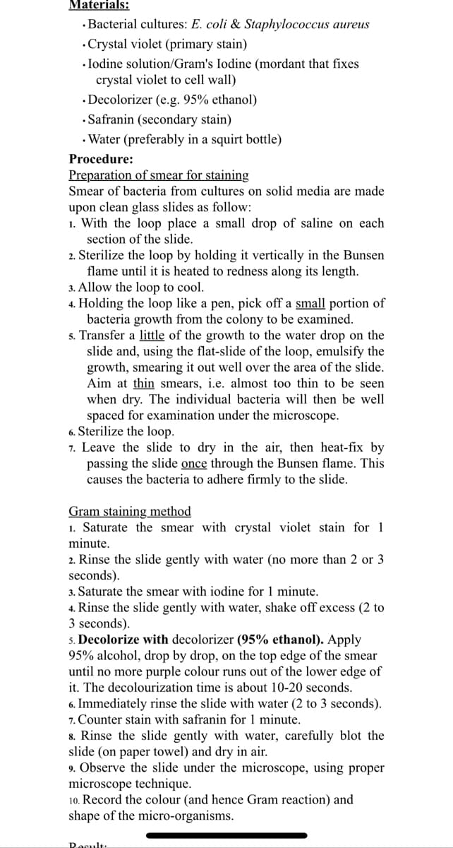
Materials: .Bacterial cultures: E. coli & Staphylococcus aureus • Crystal violet (primary stain) .Iodine solution/Gram's Iodine (mordant that fixes crystal violet to cell wall) Decolorizer (e.g. 95% ethanol) .Safranin (secondary stain) • Water (preferably in a squirt bottle) Procedure: Preparation of smear for staining Smear of bacteria from cultures on solid media are made upon clean glass slides as follow: 1. With the loop place a small drop of saline on each section of the slide. 2. Sterilize the loop by holding it vertically in the Bunsen flame until it is heated to redness along its length. 3. Allow the loop to cool. 4. Holding the loop like a pen, pick off a small portion of bacteria growth from the colony to be examined. 5. Transfer a little of the growth to the water drop on the slide and, using the flat-slide of the loop, emulsify the growth, smearing it out well over the area of the slide. Aim at thin smears, i.e. almost too thin to be seen when dry. The individual bacteria will then be well spaced for examination under the microscope. 6. Sterilize the loop. 7. Leave the slide to dry in the air, then heat-fix by passing the slide once through the Bunsen flame. This causes the bacteria to adhere firmly to the slide. Gram staining method 1. Saturate the smear with crystal violet stain for 1 minute. 2. Rinse the slide gently with water (no more than 2 or 3 seconds). 3. Saturate the smear with iodine for 1 minute. 4. Rinse the slide gently with water, shake off excess (2 to 3 seconds). 5. Decolorize with decolorizer (95% ethanol). Apply 95% alcohol, drop by drop, on the top edge of the smear until no more purple colour runs out of the lower edge of it. The decolourization time is about 10-20 seconds. 6. Immediately rinse the slide with water (2 to 3 seconds). 7. Counter stain with safranin for 1 minute. 8. Rinse the slide gently with water, carefully blot the slide (on paper towel) and dry in air. 9. Observe the slide under the microscope, using proper microscope technique. 10. Record the colour (and hence Gram reaction) and shape of the micro-organisms. Result:
Materials: .Bacterial cultures: E. coli & Staphylococcus aureus • Crystal violet (primary stain) .Iodine solution/Gram's Iodine (mordant that fixes crystal violet to cell wall) Decolorizer (e.g. 95% ethanol) .Safranin (secondary stain) • Water (preferably in a squirt bottle) Procedure: Preparation of smear for staining Smear of bacteria from cultures on solid media are made upon clean glass slides as follow: 1. With the loop place a small drop of saline on each section of the slide. 2. Sterilize the loop by holding it vertically in the Bunsen flame until it is heated to redness along its length. 3. Allow the loop to cool. 4. Holding the loop like a pen, pick off a small portion of bacteria growth from the colony to be examined. 5. Transfer a little of the growth to the water drop on the slide and, using the flat-slide of the loop, emulsify the growth, smearing it out well over the area of the slide. Aim at thin smears, i.e. almost too thin to be seen when dry. The individual bacteria will then be well spaced for examination under the microscope. 6. Sterilize the loop. 7. Leave the slide to dry in the air, then heat-fix by passing the slide once through the Bunsen flame. This causes the bacteria to adhere firmly to the slide. Gram staining method 1. Saturate the smear with crystal violet stain for 1 minute. 2. Rinse the slide gently with water (no more than 2 or 3 seconds). 3. Saturate the smear with iodine for 1 minute. 4. Rinse the slide gently with water, shake off excess (2 to 3 seconds). 5. Decolorize with decolorizer (95% ethanol). Apply 95% alcohol, drop by drop, on the top edge of the smear until no more purple colour runs out of the lower edge of it. The decolourization time is about 10-20 seconds. 6. Immediately rinse the slide with water (2 to 3 seconds). 7. Counter stain with safranin for 1 minute. 8. Rinse the slide gently with water, carefully blot the slide (on paper towel) and dry in air. 9. Observe the slide under the microscope, using proper microscope technique. 10. Record the colour (and hence Gram reaction) and shape of the micro-organisms. Result:

Human Anatomy & Physiology (11th Edition)
11th Edition
ISBN:9780134580999
Author:Elaine N. Marieb, Katja N. Hoehn
Publisher:Elaine N. Marieb, Katja N. Hoehn
Chapter1: The Human Body: An Orientation
Section: Chapter Questions
Problem 1RQ: The correct sequence of levels forming the structural hierarchy is A. (a) organ, organ system,...

Related questions

Question
For a laboratory exercise of basic techniques in microbiology : Gram stain and Microscopy.
What is the answer of table
Transcribed Image Text:Materials:
.Bacterial cultures: E. coli & Staphylococcus aureus
Crystal violet (primary stain)
.Iodine solution/Gram's Iodine (mordant that fixes
crystal violet to cell wall)
Decolorizer (e.g. 95% ethanol)
Safranin (secondary stain)
• Water (preferably in a squirt bottle)
Procedure:
Preparation of smear for staining
Smear of bacteria from cultures on solid media are made
upon clean glass slides as follow:
1. With the loop place a small drop of saline on each
section of the slide.
2. Sterilize the loop by holding it vertically in the Bunsen
flame until it is heated to redness along its length.
3. Allow the loop to cool.
4. Holding the loop like a pen, pick off a small portion of
bacteria growth from the colony to be examined.
5. Transfer a little of the growth to the water drop on the
slide and, using the flat-slide of the loop, emulsify the
growth, smearing it out well over the area of the slide.
Aim at thin smears, i.e. almost too thin to be seen
when dry. The individual bacteria will then be well
spaced for examination under the microscope.
6. Sterilize the loop.
7. Leave the slide to dry in the air, then heat-fix by
passing the slide once through the Bunsen flame. This
causes the bacteria to adhere firmly to the slide.
Gram staining method
1. Saturate the smear with crystal violet stain for 1
minute.
2. Rinse the slide gently with water (no more than 2 or 3
seconds).
3. Saturate the smear with iodine for 1 minute.
4. Rinse the slide gently with water, shake off excess (2 to
3 seconds).
5. Decolorize with decolorizer (95% ethanol). Apply
95% alcohol, drop by drop, on the top edge of the smear
until no more purple colour runs out of the lower edge of
it. The decolourization time is about 10-20 seconds.
6. Immediately rinse the slide with water (2 to 3 seconds).
7. Counter stain with safranin for 1 minute.
8. Rinse the slide gently with water, carefully blot the
slide (on paper towel) and dry in air.
9. Observe the slide under the microscope, using proper
microscope technique.
10. Record the colour (and hence Gram reaction) and
shape of the micro-organisms.
Result:
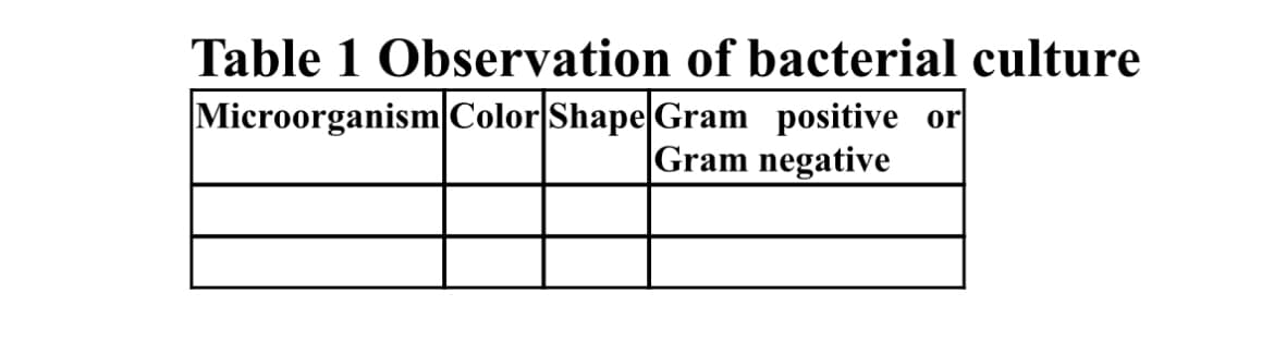
Transcribed Image Text:Table 1 Observation of bacterial culture
Microorganism Color Shape Gram positive or
Gram negative
Expert Solution
This question has been solved!
Explore an expertly crafted, step-by-step solution for a thorough understanding of key concepts.

Step by step
Solved in 3 steps

Knowledge Booster


Learn more about
Need a deep-dive on the concept behind this application? Look no further. Learn more about this topic, biology and related others by exploring similar questions and additional content below.Recommended textbooks for you
Human Anatomy & Physiology (11th Edition)
Biology
ISBN:
9780134580999
Author:
Elaine N. Marieb, Katja N. Hoehn
Publisher:
PEARSON

Biology 2e
Biology
ISBN:
9781947172517
Author:
Matthew Douglas, Jung Choi, Mary Ann Clark
Publisher:
OpenStax

Anatomy & Physiology
Biology
ISBN:
9781259398629
Author:
McKinley, Michael P., O'loughlin, Valerie Dean, Bidle, Theresa Stouter
Publisher:
Mcgraw Hill Education,
Human Anatomy & Physiology (11th Edition)
Biology
ISBN:
9780134580999
Author:
Elaine N. Marieb, Katja N. Hoehn
Publisher:
PEARSON
Biology 2e
Biology
ISBN:
9781947172517
Author:
Matthew Douglas, Jung Choi, Mary Ann Clark
Publisher:
OpenStax
Anatomy & Physiology
Biology
ISBN:
9781259398629
Author:
McKinley, Michael P., O'loughlin, Valerie Dean, Bidle, Theresa Stouter
Publisher:
Mcgraw Hill Education,

Molecular Biology of the Cell (Sixth Edition)
Biology
ISBN:
9780815344322
Author:
Bruce Alberts, Alexander D. Johnson, Julian Lewis, David Morgan, Martin Raff, Keith Roberts, Peter Walter
Publisher:
W. W. Norton & Company

Laboratory Manual For Human Anatomy & Physiology
Biology
ISBN:
9781260159363
Author:
Martin, Terry R., Prentice-craver, Cynthia
Publisher:
McGraw-Hill Publishing Co.

Inquiry Into Life (16th Edition)
Biology
ISBN:
9781260231700
Author:
Sylvia S. Mader, Michael Windelspecht
Publisher:
McGraw Hill Education