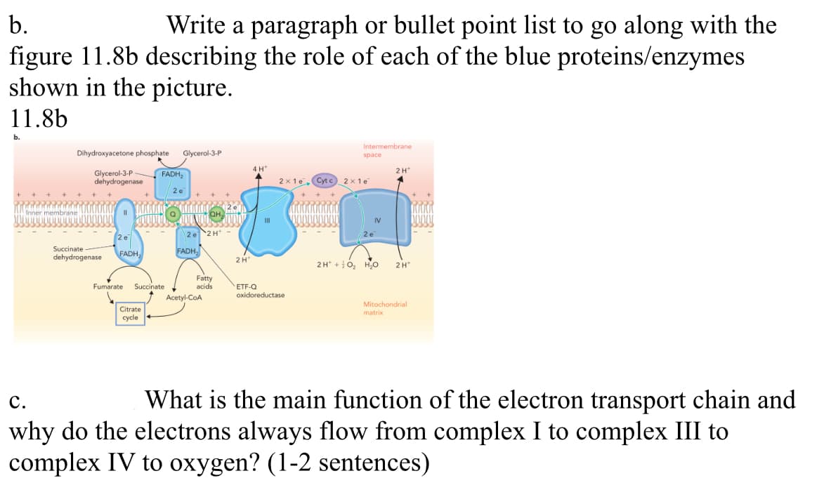
b. Write a paragraph or bullet point list to go along with the figure 11.8b describing the role of each of the blue proteins/enzymes shown in the picture. 11.8b b. Dihydroxyacetone phosphate Glycerol-3-P Glycerol-3-P dehydrogenase Succinate dehydrogenase KAKOR 1000000 JOLANTOLA FADH, FADH₂ Fumarate Succinate Citrate cycle 2e 2H- FADH₂ Fatty acids Acetyl-CoA 2H 4 H+ 111 Intermembrane ETF-Q oxidoreductase space 2x1e Cytc 2x1e 2 e 2H+O₂ H₂O 2 H matrix 2 H Mitochondrial C. What is the main function of the electron transport chain and why do the electrons always flow from complex I to complex III to complex IV to oxygen? (1-2 sentences)
b. Write a paragraph or bullet point list to go along with the figure 11.8b describing the role of each of the blue proteins/enzymes shown in the picture. 11.8b b. Dihydroxyacetone phosphate Glycerol-3-P Glycerol-3-P dehydrogenase Succinate dehydrogenase KAKOR 1000000 JOLANTOLA FADH, FADH₂ Fumarate Succinate Citrate cycle 2e 2H- FADH₂ Fatty acids Acetyl-CoA 2H 4 H+ 111 Intermembrane ETF-Q oxidoreductase space 2x1e Cytc 2x1e 2 e 2H+O₂ H₂O 2 H matrix 2 H Mitochondrial C. What is the main function of the electron transport chain and why do the electrons always flow from complex I to complex III to complex IV to oxygen? (1-2 sentences)

Biochemistry
6th Edition
ISBN:9781305577206
Author:Reginald H. Garrett, Charles M. Grisham
Publisher:Reginald H. Garrett, Charles M. Grisham
Chapter20: Electron Transport And Oxidative Phosphorylation
Section: Chapter Questions
Problem 11P

Related questions
Question
This is Biochemistry, there is a max of 3 parts for the question due to guidelines. Please answer each section to the best of your ability. Please provide thorough and clear work with answers. Thank you.
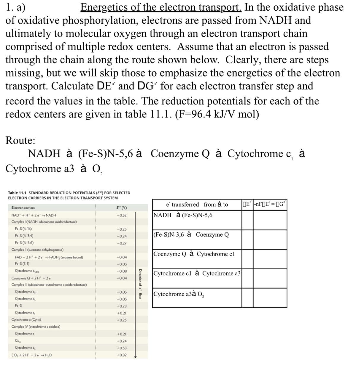
Transcribed Image Text:1. a)
Energetics of the electron transport. In the oxidative phase
of oxidative phosphorylation, electrons are passed from NADH and
ultimately to molecular oxygen through an electron transport chain
comprised of multiple redox centers. Assume that an electron is passed
through the chain along the route shown below. Clearly, there are steps
missing, but we will skip those to emphasize the energetics of the electron
transport. Calculate DE° and DG" for each electron transfer step and
record the values in the table. The reduction potentials for each of the
redox centers are given in table 11.1. (F=96.4 kJ/V mol)
Route:
1
NADH à (Fe-S)N-5,6 à Coenzyme Q à Cytochrome c, à
Cytochrome a3 à O,
Table 11.1 STANDARD REDUCTION POTENTIALS (E°') FOR SELECTED
ELECTRON CARRIERS IN THE ELECTRON TRANSPORT SYSTEM
Electron carriers
NAD + H+ +2e → NADH
Complex I (NADH-ubiquinone oxidoreductase)
Fe-S (N-1b)
Fe-S (N-3.4)
Fe-S (N-5,6)
Complex II (succinate dehydrogenase)
FAD + 2 H+2e → FADH₂ (enzyme bound)
Fe-S (S-1)
Cytochrome b560
Coenzyme Q + 2H+ + 2 e
Complex III (ubiquinone-cytochrome c oxidoreductase)
Cytochrome b
Cytochrome b
Fe-S
Cytochrome c₁
Cytochrome c (Cyt c)
Complex IV (cytochrome c oxidase)
Cytochrome a
CUA
Cytochrome a
O₂+ 2H+2e →H₂O
E" (V)
-0.32
-0.25
-0.24
-0.27
-0.04
-0.03
-0.08
+0.04
+0.03
-0.03
+0.28
+0.21
+0.23
+0.21
+0.24
+0.38
+0.82
Direction of e flow
e transferred from à to
NADH à (Fe-S)N-5,6
(Fe-S)N-3,6 à Coenzyme Q
Coenzyme Q à Cytochrome cl
Cytochrome cl à Cytochrome a3|
Cytochrome a3à 02
E-nF E=GⓇ
Expert Solution
This question has been solved!
Explore an expertly crafted, step-by-step solution for a thorough understanding of key concepts.

This is a popular solution!

Trending now
This is a popular solution!

Step by step
Solved in 3 steps with 26 images

Recommended textbooks for you
Biochemistry
Biochemistry
ISBN:
9781305577206
Author:
Reginald H. Garrett, Charles M. Grisham
Publisher:
Cengage Learning
Biochemistry
Biochemistry
ISBN:
9781305577206
Author:
Reginald H. Garrett, Charles M. Grisham
Publisher:
Cengage Learning