SIZE ESTIAMTION- Since we know the diameter of the field of view, we can estimate the size of a specimen by determining the proportion of the field of view the object (cell) takes up. Based on the diameters in um above, estimate the size of the following object as if looking through the 1. low power 2. high power 3. oil
SIZE ESTIAMTION- Since we know the diameter of the field of view, we can estimate the size of a specimen by determining the proportion of the field of view the object (cell) takes up. Based on the diameters in um above, estimate the size of the following object as if looking through the 1. low power 2. high power 3. oil

Biology 2e
2nd Edition
ISBN:9781947172517
Author:Matthew Douglas, Jung Choi, Mary Ann Clark
Publisher:Matthew Douglas, Jung Choi, Mary Ann Clark
Chapter4: Cell Structure
Section: Chapter Questions
Problem 25CTQ: In your everyday life, you have probably noticed that certain instruments are ideal for certain...

Related questions
Question
I need the answer as soon as possible
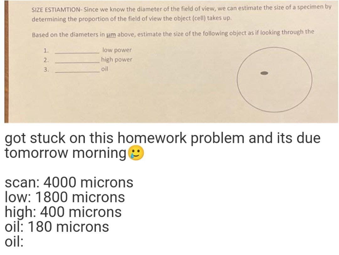
Transcribed Image Text:SIZE ESTIAMTION- Since we know the diameter of the field of view, we can estimate the size of a specimen by
determining the proportion of the field of view the object (cell) takes up.
Based on the diameters in um above, estimate the size of the following object as if looking through the
1.
low power
2.
high power
3.
oil
got stuck on this homework problem and its due
tomorrow morning
scan: 4000 microns
low: 1800 microns
high: 400 microns
oil: 180 microns
oil:
Expert Solution
This question has been solved!
Explore an expertly crafted, step-by-step solution for a thorough understanding of key concepts.

Step by step
Solved in 2 steps with 4 images

Knowledge Booster


Learn more about
Need a deep-dive on the concept behind this application? Look no further. Learn more about this topic, biology and related others by exploring similar questions and additional content below.Recommended textbooks for you
Biology 2e
Biology
ISBN:
9781947172517
Author:
Matthew Douglas, Jung Choi, Mary Ann Clark
Publisher:
OpenStax

Biology 2e
Biology
ISBN:
9781947172517
Author:
Matthew Douglas, Jung Choi, Mary Ann Clark
Publisher:
OpenStax