Cytokinesis , or division of the cytoplasm to create two new daughter cells , occurs in conjunction with telophase . Examine the telophase micrographs of the onion root tip and whitefish blastodisc cells . How does cytokinesis look different for these two cells ? Explain why cytokinesis looks different between those cells
Cytokinesis , or division of the cytoplasm to create two new daughter cells , occurs in conjunction with telophase . Examine the telophase micrographs of the onion root tip and whitefish blastodisc cells . How does cytokinesis look different for these two cells ? Explain why cytokinesis looks different between those cells

Biochemistry
6th Edition
ISBN:9781305577206
Author:Reginald H. Garrett, Charles M. Grisham
Publisher:Reginald H. Garrett, Charles M. Grisham
Chapter28: Dna Metabolism: Replication, Recombination, And Repair
Section: Chapter Questions
Problem 19P: Figure 28.11 depicts the eukaryotic cell cycle. Many cell types “exit� the cell cycle and...

Related questions
Question
Cytokinesis , or division of the cytoplasm to create two new daughter cells , occurs in conjunction with telophase . Examine the telophase micrographs of the onion root tip and whitefish blastodisc cells . How does cytokinesis look different for these two cells ? Explain why cytokinesis looks different between those cells
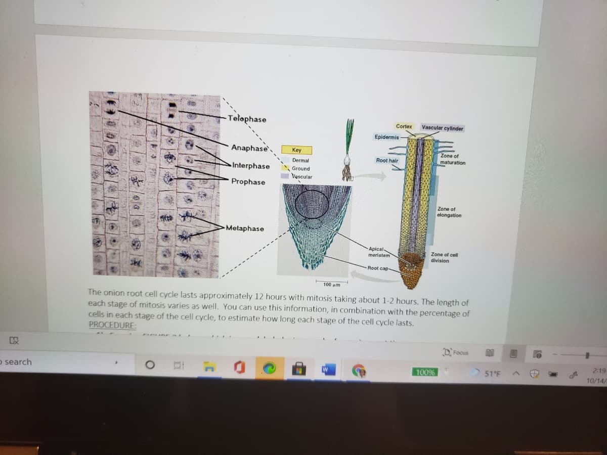
Transcribed Image Text:Telophase
Cortex
Vascular cylinder
Epidermis
Anaphase
Key
Zone of
maturation
Dermal
Root hair
Interphase
Ground
Vascular
Prophase
Zone of
elongation
Metaphase
Apical
meristem
Zone of cell
division
Root cap-
100 um
The onion root cell cycle lasts approximately 12 hours with mitosis taking about 1-2 hours. The length of
each stage of mitosis varies as well. You can use this information, in combination with the percentage of
cells in each stage of the cell cycle, to estimate how long each stage of the cell cycle lasts.
PROCEDURE:
O, Focus
o search
100%
51°F
2:19
10/14/
1 |ま113|)
Transcribed Image Text:MITOSIS ASSIGNMENT - Saved to this PC
O Search
christine
Design
Layout
References
Mailings
Review
View
Help
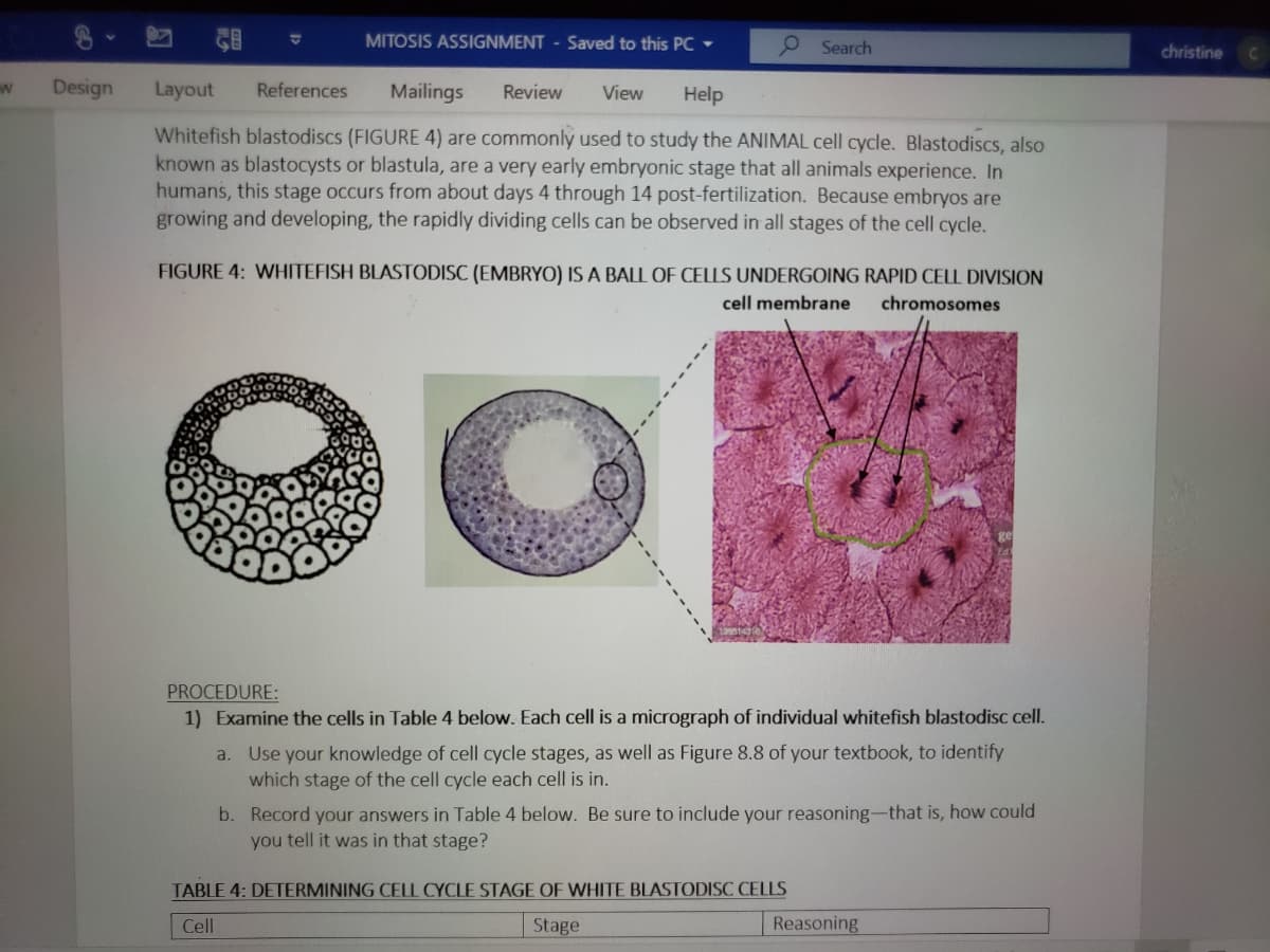
Whitefish blastodiscs (FIGURE 4) are commonly used to study the ANIMAL cell cycle. Blastodiscs, also
known as blastocysts or blastula, are a very early embryonic stage that all animals experience. In
humans, this stage occurs from about days 4 through 14 post-fertilization. Because embryos are
growing and developing, the rapidly dividing cells can be observed in all stages of the cell cycle.
FIGURE 4: WHITEFISH BLASTODISC (EMBRYO) IS A BALL OF CELLS UNDERGOING RAPID CELL DIVISION
cell membrane
chromosomes
PROCEDURE:
1) Examine the cells in Table 4 below. Each cell is a micrograph of individual whitefish blastodisc cell.
a. Use your knowledge of cell cycle stages, as well as Figure 8.8 of your textbook, to identify
which stage of the cell cycle each cell is in.
b. Record your answers in Table 4 below. Be sure to include your reasoning-that is, how could
you tell it was in that stage?
TABLE 4: DETERMINING CELL CYCLE STAGE OF WHITE BLASTODISC CELLS
Cell
Stage
Reasoning
Expert Solution
This question has been solved!
Explore an expertly crafted, step-by-step solution for a thorough understanding of key concepts.

This is a popular solution!

Trending now
This is a popular solution!

Step by step
Solved in 2 steps

Recommended textbooks for you
Biochemistry
Biochemistry
ISBN:
9781305577206
Author:
Reginald H. Garrett, Charles M. Grisham
Publisher:
Cengage Learning

Biology 2e
Biology
ISBN:
9781947172517
Author:
Matthew Douglas, Jung Choi, Mary Ann Clark
Publisher:
OpenStax
Biochemistry
Biochemistry
ISBN:
9781305577206
Author:
Reginald H. Garrett, Charles M. Grisham
Publisher:
Cengage Learning
Biology 2e
Biology
ISBN:
9781947172517
Author:
Matthew Douglas, Jung Choi, Mary Ann Clark
Publisher:
OpenStax