Focusing on the mechanism linking complex I and ATP synthase depicted in figure 3 in the article, compare that hypothetical mechanism to the classical presentation described in textbooks. What are the major differences between this mechanism and Peter Mitchel’s original chemiosmotic theory? What are the similarities.
Focusing on the mechanism linking complex I and ATP synthase depicted in figure 3 in the article, compare that hypothetical mechanism to the classical presentation described in textbooks. What are the major differences between this mechanism and Peter Mitchel’s original chemiosmotic theory? What are the similarities.

Biochemistry
6th Edition
ISBN:9781305577206
Author:Reginald H. Garrett, Charles M. Grisham
Publisher:Reginald H. Garrett, Charles M. Grisham
Chapter27: Metabolic Integration And Organ Specialization
Section: Chapter Questions
Problem 3P

Related questions
Question
Focusing on the mechanism linking complex I and ATP synthase depicted in figure 3 in the article, compare that hypothetical mechanism to the classical presentation described in textbooks. What are the major differences between this mechanism and Peter Mitchel’s original chemiosmotic theory? What are the similarities.
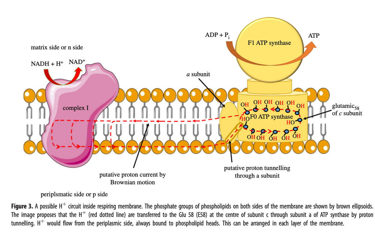
Transcribed Image Text:matrix side or n side
NADH + H+ NAD+
complex I
U: U
DESODO
putative proton current by
Brownian motion
ADP + Pi
a subunit
OG
F1 ATP synthase
OH
ОН ОН ОН ОН
do < od
HOFO ATP synthase
HO
ОН ОН ОН ОН ОН
putative proton tunnelling
through a subunit
OH
ATP
00
glutamic 58
of c subunit
periplsmatic side or p side
Figure 3. A possible H+ circuit inside respiring membrane. The phosphate groups of phospholipids on both sides of the membrane are shown by brown ellipsoids.
The image proposes that the H+ (red dotted line) are transferred to the Glu 58 (E58) at the centre of subunit c through subunit a of ATP synthase by proton
tunnelling. H+ would flow from the periplasmic side, always bound to phospholipid heads. This can be arranged in each layer of the membrane.
Expert Solution
This question has been solved!
Explore an expertly crafted, step-by-step solution for a thorough understanding of key concepts.

This is a popular solution!

Trending now
This is a popular solution!

Step by step
Solved in 4 steps

Recommended textbooks for you
Biochemistry
Biochemistry
ISBN:
9781305577206
Author:
Reginald H. Garrett, Charles M. Grisham
Publisher:
Cengage Learning
Biochemistry
Biochemistry
ISBN:
9781305577206
Author:
Reginald H. Garrett, Charles M. Grisham
Publisher:
Cengage Learning