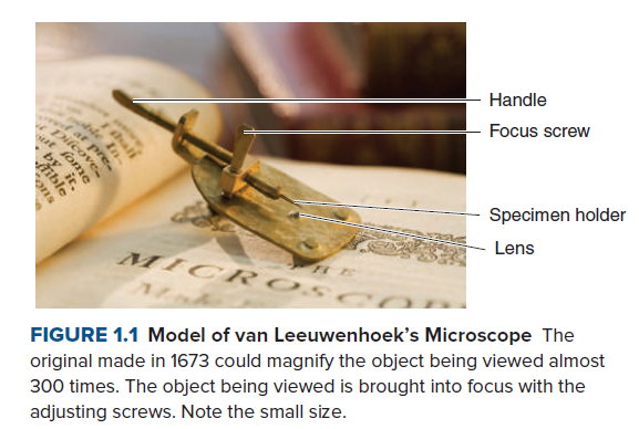
wwwat pre. Handle Focus screw le In Dilcove Specimen holder Lens MICR 0S FIGURE 1.1 Model of van Leeuwenhoek's Microscope The original made in 1673 could magnify the object being viewed almost 300 times. The object being viewed is brought into focus with the adjusting screws. Note the small size. hali by it, Aible


Microscopic examination
The analysis of minute organisms, cellular organization of any biological structure, and composition of body fluids with the help of a microscope is known as microscopic examination. The magnification of specimens or samples under study helps in attaining a clearer picture of it.
Gram Staining
Named after Hans Christian Gram, a Danish bacteriologist, Gram stain is one of the most powerful staining techniques within microbiology. This technique was introduced in 1882 to identify pneumonia-causing organisms. The Gram staining technique uses crystal violet or methylene blue as primary staining colors to distinguish gram-positive from gram-negative organisms. Under a microscope, the gram-positive organisms appear purple-brown, retaining the primary color. Gram-negative organisms appear pink or red as they do not acquire the color of the primary stain.
What kinds of organisms did van Leeuwenhoek observe though his microscope?

Step by step
Solved in 2 steps


