
Anatomy & Physiology
1st Edition
ISBN:9781938168130
Author:Kelly A. Young, James A. Wise, Peter DeSaix, Dean H. Kruse, Brandon Poe, Eddie Johnson, Jody E. Johnson, Oksana Korol, J. Gordon Betts, Mark Womble
Publisher:Kelly A. Young, James A. Wise, Peter DeSaix, Dean H. Kruse, Brandon Poe, Eddie Johnson, Jody E. Johnson, Oksana Korol, J. Gordon Betts, Mark Womble
Chapter23: The Digestive System
Section: Chapter Questions
Problem 16RQ: Which of these statements about the pharynx is tine? It extends horn the nasal and oral cavities...

Related questions

Question
read the article that is in the images and write the most important data
Transcribed Image Text:11:52 p. m. Mar 28 mar.
198
PART TWO Radiographic Positioning and Related Anatomy
C
RtL B
web.whatsapp.com
D
LeL
VD
DV
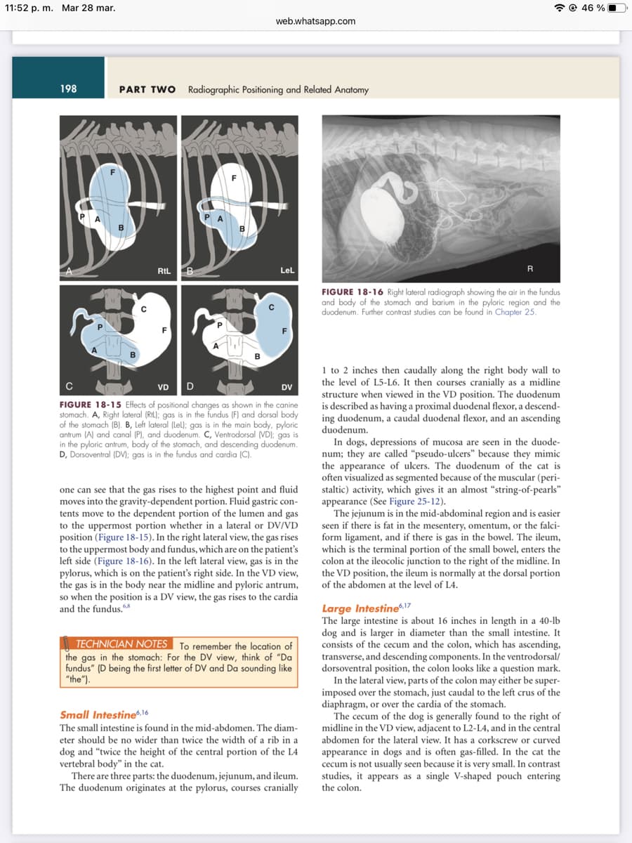
FIGURE 18-15 Effects of positional changes as shown in the canine
stomach. A, Right lateral (RL); gas is in the fundus (F) and dorsal body
of the stomach (B). B, Left lateral (lel); gas is in the main body, pyloric
antrum (A) and canal (P), and duodenum. C, Ventrodorsal (VD); gas is
in the pyloric antrum, body of the stomach, and descending duodenum.
D, Dorsoventral (DV); gas is in the fundus and cardia (C).
one can see that the gas rises to the highest point and fluid
moves into the gravity-dependent portion. Fluid gastric con-
tents move to the dependent portion of the lumen and gas
to the uppermost portion whether in a lateral or DV/VD
position (Figure 18-15). In the right lateral view, the gas rises
to the uppermost body and fundus, which are on the patient's
left side (Figure 18-16). In the left lateral view, gas is in the
pylorus, which on the patient's right side. In the VD view,
the gas is in the body near the midline and pyloric antrum,
so when the position is a DV view, the gas rises to the cardia
and the fundus.68
TECHNICIAN NOTES To remember the location of
the gas in the stomach: For the DV view, think of "Da
fundus" (D being the first letter of DV and Da sounding like
"the").
Small Intestine6,16
The small intestine is found in the mid-abdomen. The diam-
eter should be no wider than twice the width of a rib in a
dog and "twice the height of the central portion of the L4
vertebral body" in the cat.
There are three parts: the duodenum, jejunum, and ileum.
The duodenum originates at the pylorus, courses cranially
R
FIGURE 18-16 Right lateral radiograph showing the air in the fundus
and body of the stomach and barium in the pyloric region and the
duodenum. Further contrast studies can be found in Chapter 25.
1 to 2 inches then caudally along the right body wall to
the level of L5-L6. It then courses cranially as a midline
structure when viewed in the VD position. The duodenum
is described as having a proximal duodenal flexor, a descend-
ing duodenum, a caudal duodenal flexor, and an ascending
duodenum.
In dogs, depressions of mucosa are seen in the duode-
num; they are called "pseudo-ulcers" because they mimic
the appearance of ulcers. The duodenum of the cat is
often visualized as segmented because of the muscular (peri-
staltic) activity, which gives it an almost "string-of-pearls"
appearance (See Figure 25-12).
The jejunum is in the mid-abdominal region and is easier
seen if there is fat in the mesentery, omentum, or the falci-
form ligament, and if there is gas in the bowel. The ileum,
which is the terminal portion of the small bowel, enters the
colon at the ileocolic junction to the right of the midline. In
the VD position, the ileum is normally at the dorsal portion.
of the abdomen at the level of L4.
Large Intestine6,17
The large intestine is about 16 inches in length in a 40-lb
dog and is larger in diameter than the small intestine. It
consists of the cecum and the colon, which has ascending,
transverse, and descending components. In the ventrodorsal/
dorsoventral position, the colon looks like a question mark.
In the lateral view, parts of the colon may either be super-
imposed over the stomach, just caudal to the left crus of the
diaphragm, or over the cardia of the stomach.
The cecum of the dog is generally found to the right of
midline in the VD view, adjacent to L2-L4, and in the central
abdomen for the lateral view. It has a corkscrew or curved
appearance in dogs and is often gas-filled. In the cat the
cecum is not usually seen because it is very small. In contrast
studies, it appears as a single V-shaped pouch entering
the colon.
46 %
Transcribed Image Text:11:52 p. m. Mar 28 mar.
U Blackboard...
260 de 633
xblob:https://...
web.whatsapp.com
Tips for mas...
The ascending colon is to the right of midline and is
found dorsally. The ascending colon is often indented along
the medial wall adjacent to the cecum, at the site where the
ileum enters the ascending colon. This is known as the ileo-
colonic sphincter. The transverse colon appears circular in
configuration and lies caudal to the stomach. In the lateral
view, the transverse colon can often be mistaken for a mass
lesion. The descending colon generally lies adjacent to the
left body wall, but it may change position to either the
midline or the right side in the mid- and distal regions,
especially if the animal is obese, the intestine is full of fecal
matter, or the urinary bladder is distended.
The rectum is located in the pelvic canal.
Pancreas 6,7
ormal pancreas is visualized radiographically. Even
pancreatitis may not show any radiographic abnormalities
but may cause organ displacements. The right limb of the
pancreas lies adjacent and caudal to the caudal margin of
the stomach. The left portion is medial to and beside the
descending duodenum. Ultrasonography is a better tool to
identify a diseased pancreas.
Evaluation of the Urogenital System,
Adrenal Glands, and Abdominal
Lymph Nodes
Kidneys6,11
The kidneys are soft-tissue opacities found in the retroperi-
toneal space. Canine kidneys are elongated, and feline
kidneys are short and round. In the dog the right kidney is
usually found between T12 and L2, and in the cat between
L1 and L3. The left kidney position, which can be more vari-
able, is between L2 and L4 in the dog and L3 and L5 in the
cat. Normal kidney size is generally judged in the VD view
with the use of L2 as a comparison. In the dog, the normal
kidney should be 2½ to 3½ times the length of L2, and in
the cat, 2 to 3 times. However, this observation is a guideline
only, because there are variations with factors such as age,
size, sex and blood pressure. The kidneys may be displaced
caudally by a full stomach or cranially by an enlarged uterus.
Both kidneys are usually identifiable on the feline radio-
graph. The left kidney is seen in most dogs with normal
body condition, but only the caudal pole of the right kidney
can be seen on a lateral radiograph and the organ is
usually not seen at all in the VD radiograph. On the right
lateral radiograph, the left kidney will be displaced from its
dorsal position; it may be in the mid-abdomen and is often
more visible than on a left lateral radiograph, especially in
cats and obese dogs, in which there is more perirenal and
retroperitoneal fat.
The margins of the kidneys should be smooth and of
homogenous soft-tissue opacity. If a unilateral renal lesion
is suspected, place the abnormal kidney in the upper posi-
tion. It will be more "bean-shaped" because the upper kidney
rotates on the longitudinal axis. This rotation allows that
region, which is well covered with fat, to be presented.
G importancia...
nurse Take a Bite...
CHAPTER 18 Small Animal Abdomen
+
ódulos / C... ogía o...
199
tangentially to the x-ray beam. The increased object-film
distance makes it appear more magnified as well.
Adrenal Glands6,7
Normal adrenal glands are not generally visualized on the
radiograph, being better viewed on ultrasound and CT scan-
ning. They are located in the retroperitoneal space just cra-
niomedial to each kidney.
Urinary Bladder6,12
The urinary bladder is found in the caudoventral abdomen
ventral to the rectum and descending colon. In the dog, the
caudally facing neck of this pear-shaped organ is cranial to
the pubis. In the cat, the bladder is more rounded and slightly
more cranial.
cra-
A distended bladder may displace abdomina
nially and the descending colon to right. An empty urinary
bladder may not be visible, but if it contains urine, it may
be seen on either the left or right side or centered on the
midline.
Uterus and Ovaries¹0
The uterus and ovaries are not normally visualized on survey
radiographs, although the body of the uterus may sometimes
be visible between the bladder neck and colon in obese
animals. The uterus is usually visualized only if it is abnor-
mally large or gravid. Fetal skeletons become visible at
approximately 42- to 45 days of gestation.
Urethra and Ureters6,11,13
Neither the urethra or the ureters are visible on a radiograph
without contrast media. The female urethra is shorter and
wider than that of the male. The male urethra is long and
thin. The canine male urethra consists of three parts while
the feline urethra has two parts. The ureters are in the retro-
peritoneal space and if injected with contrast media should
not be larger than 2 to 3 mm.
Prostate Gland and Testicles6,14,15
The prostate is found in the pelvic canal and is not visible
radiographically in the cat. In older dogs it may lie within
the abdomen, as it may in young dogs when pulled cranially
by a full urinary bladder. The prostate diameter in the dog
should be no greater than two-thirds the width of the pelvic
inlet on the VD view. The testicles are not normally evalu-
ated with conventional radiography.
Abdominal Lymph Nodes
Abdominal lymph nodes are not normally visualized but
may be seen if there is a considerable increase in size or a
change in opacity. Ultrasound is an excellent modality to
evaluate lymph nodes (see the description in Chapter 13).
46%
NOTE
For information on alternate modes of imaging, please see
Sections 3 and 4 in Part One. Further images available on line.
Expert Solution
This question has been solved!
Explore an expertly crafted, step-by-step solution for a thorough understanding of key concepts.

This is a popular solution!

Trending now
This is a popular solution!

Step by step
Solved in 2 steps

Knowledge Booster


Learn more about
Need a deep-dive on the concept behind this application? Look no further. Learn more about this topic, biology and related others by exploring similar questions and additional content below.Recommended textbooks for you
Anatomy & Physiology
Biology
ISBN:
9781938168130
Author:
Kelly A. Young, James A. Wise, Peter DeSaix, Dean H. Kruse, Brandon Poe, Eddie Johnson, Jody E. Johnson, Oksana Korol, J. Gordon Betts, Mark Womble
Publisher:
OpenStax College

Cardiopulmonary Anatomy & Physiology
Biology
ISBN:
9781337794909
Author:
Des Jardins, Terry.
Publisher:
Cengage Learning,

Surgical Tech For Surgical Tech Pos Care
Health & Nutrition
ISBN:
9781337648868
Author:
Association
Publisher:
Cengage
Anatomy & Physiology
Biology
ISBN:
9781938168130
Author:
Kelly A. Young, James A. Wise, Peter DeSaix, Dean H. Kruse, Brandon Poe, Eddie Johnson, Jody E. Johnson, Oksana Korol, J. Gordon Betts, Mark Womble
Publisher:
OpenStax College
Cardiopulmonary Anatomy & Physiology
Biology
ISBN:
9781337794909
Author:
Des Jardins, Terry.
Publisher:
Cengage Learning,
Surgical Tech For Surgical Tech Pos Care
Health & Nutrition
ISBN:
9781337648868
Author:
Association
Publisher:
Cengage

Comprehensive Medical Assisting: Administrative a…
Nursing
ISBN:
9781305964792
Author:
Wilburta Q. Lindh, Carol D. Tamparo, Barbara M. Dahl, Julie Morris, Cindy Correa
Publisher:
Cengage Learning

Fundamentals of Sectional Anatomy: An Imaging App…
Biology
ISBN:
9781133960867
Author:
Denise L. Lazo
Publisher:
Cengage Learning