Red Blood Cell: Figure 9. Changes to the beta-globin subunit of hemoglobin in sickle cell disease and the functional consequence for red blood cells. Image modified from here in accordance with Creative Commons License. Watch this video about sickle cell anemia, then answer the questions below. 3. Figure 9 illustrates the molecular cause of the most common version of sickle cell disease. A valine is substituted for a glutamate in hemoglobin in sickle cell disease. Which levels of protein structure are altered, and specifically, which types of interaction are likely to be changed in determining the shape of the protein? Explain your answer. 4. Normal red blood cells can easily travel through blood vessels, whereas sickle- shaped red blood cells get stuck. This is the basis of sickle cell anemia. What does this tell you about the relationship between the sequence of amino acids, the shape of the protein and its function in a cell/organism? mmarize what you found for each level
Red Blood Cell: Figure 9. Changes to the beta-globin subunit of hemoglobin in sickle cell disease and the functional consequence for red blood cells. Image modified from here in accordance with Creative Commons License. Watch this video about sickle cell anemia, then answer the questions below. 3. Figure 9 illustrates the molecular cause of the most common version of sickle cell disease. A valine is substituted for a glutamate in hemoglobin in sickle cell disease. Which levels of protein structure are altered, and specifically, which types of interaction are likely to be changed in determining the shape of the protein? Explain your answer. 4. Normal red blood cells can easily travel through blood vessels, whereas sickle- shaped red blood cells get stuck. This is the basis of sickle cell anemia. What does this tell you about the relationship between the sequence of amino acids, the shape of the protein and its function in a cell/organism? mmarize what you found for each level

Chapter3: Cells
Section: Chapter Questions
Problem 5C

Related questions
Question
Transcribed Image Text:Normal
Sickle CelI Disease
Partial Amino Acid
Sequence for Beta Globin: Pro-Glu-Glu
Pro Val
Glu
Hemoglobin Molecule:
Red Blood Cell:
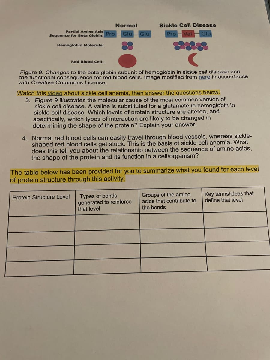
Figure 9. Changes to the beta-globin subunit of hemoglobin in sickle cell disease and
the functional consequence for red blood cells. Image modified from here in accordance
with Creative Commons License.
Watch this video about sickle cell anemia, then answer the questions below.
3. Figure 9 illustrates the molecular cause of the most common version of
sickle cell disease. A valine is substituted for a glutamate in hemoglobin in
sickle cell disease. Which levels of protein structure are altered, and
specifically, which types of interaction are likely to be changed in
determining the shape of the protein? Explain your answer.
4. Normal red blood cells can easily travel through blood vessels, whereas sickle-
shaped red blood cells get stuck. This is the basis of sickle cell anemia. What
does this tell you about the relationship between the sequence of amino acids,
the shape of the protein and its function in a cell/organism?
The table below has been provided for you to summarize what you found for each level
of protein structure through this activity.
Types of bonds
generated to reinforce
that level
Groups of the amino
acids that contribute to
Key terms/ideas that
define that level
Protein Structure Level
the bonds
Transcribed Image Text:QUATERNARY STRUCTURE
One final level of structure exists for some, but not all, proteins. This is called
quaternary structure. Proteins that have quaternary structure are formed from two or
more polypeptides that assemble into one active structure. The different polypeptides in
a protein with quaternary structure are often called subunits. These subunits may be
identical or different. One example of a protein with quaternary structure is hemoglobin,
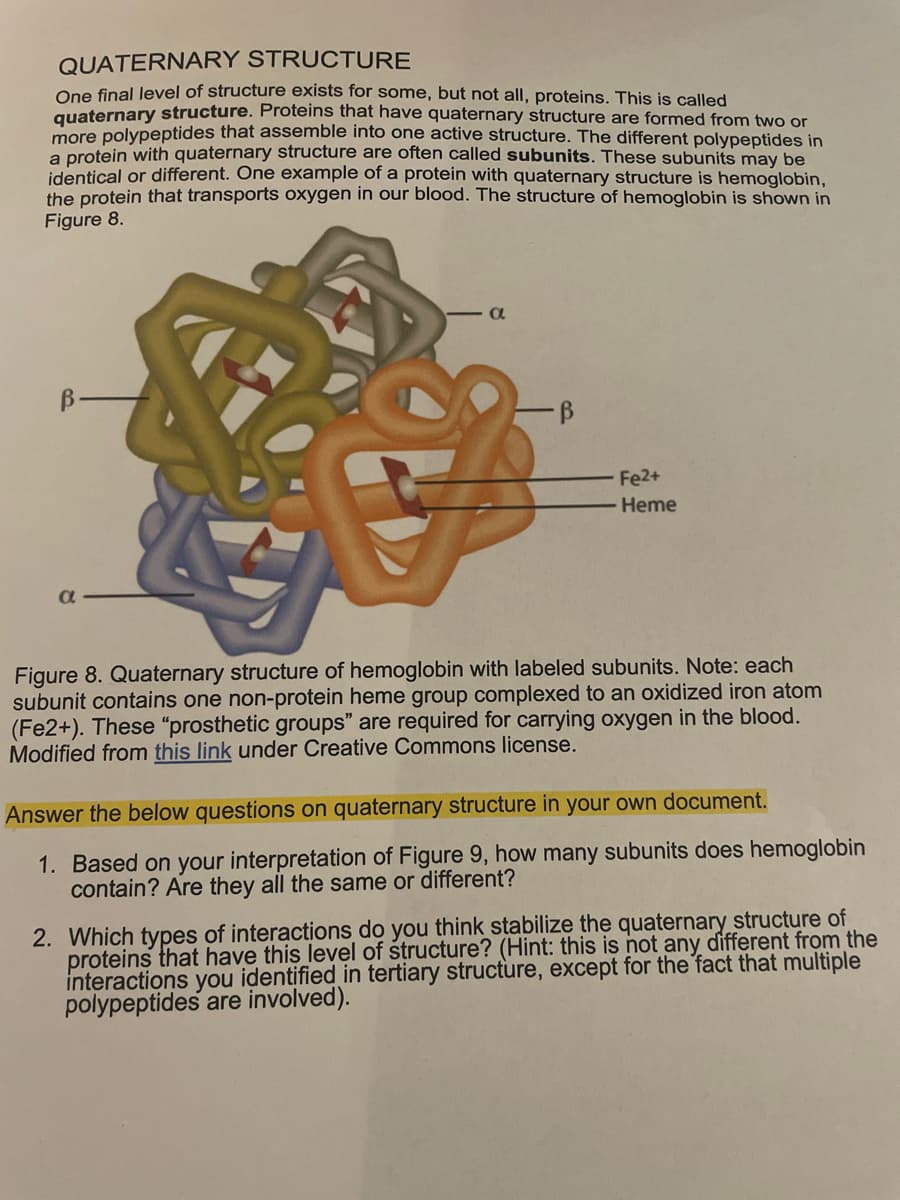
the protein that transports oxygen in our blood. The structure of hemoglobin is shown in
Figure 8.
B-
Fe2+
Heme
a
Figure 8. Quaternary structure of hemoglobin with labeled subunits. Note: each
subunit contains one non-protein heme group complexed to an oxidized iron atom
(Fe2+). These “prosthetic groups" are required for carrying oxygen in the blood.
Modified from this link under Creative Commons license.
Answer the below questions on quaternary structure in your own document.
1. Based on your interpretation of Figure 9, how many subunits does hemoglobin
contain? Are they all the same or different?
2. Which types of interactions do you think stabilize the quaternary structure of
proteins that have this level of structure? (Hint: this is not any different from the
interactions you identified in tertiary structure, except for the fact that multiple
polypeptides are involved).
Expert Solution
This question has been solved!
Explore an expertly crafted, step-by-step solution for a thorough understanding of key concepts.

This is a popular solution!

Trending now
This is a popular solution!

Step by step
Solved in 2 steps

Recommended textbooks for you