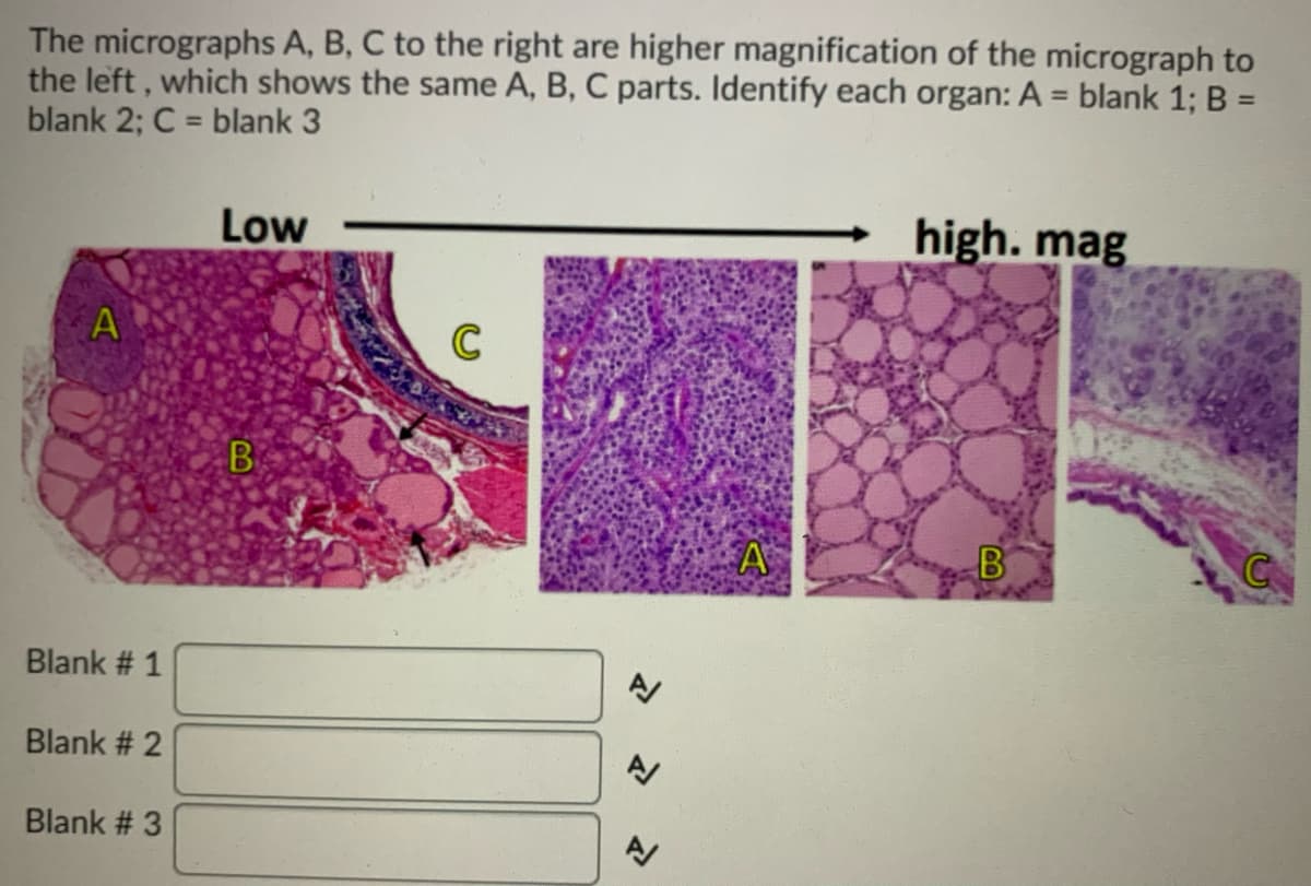
The micrographs A, B, C to the right are higher magnification of the micrograph to the left , which shows the same A, B, C parts. Identify each organ: A = blank 1; B = blank 2; C = blank 3 %3D %3D Low high. mag C B. Blank # 1 Blank # 2 Blank # 3
Q: What is staining in histology
A: Hello, thank you for your question. Since the typed question and the question in the image do not…
Q: What did you notice about the Human Palmar Skin photomicrographs shown at (a) 400x vs (b) 100x TM?…
A: Note: since you have posted multiple questions and didn’t mention which one needs to be solved. As…
Q: You have a microscope equipped with 10 x oculars and 4 objectives, which exhibit the following…
A: Objective lens The objectives lens have various magnification powers with common values of 4x, 10x,…
Q: what happens to the field of view as magnification is increased useing shccdssively higher power…
A:
Q: Imagine someone is using a microscope with 5x eye pieces (instead of 10x) Complete the sentence.…
A: In light microscopy, there are several different types of microscopes, but the four most common are…
Q: Match the terms on the left with its description or definition on the right. There is only one…
A: (Please note you have posted multiple questions. We can solve 1 question at a time. Please post rest…
Q: "Dissecting microscope provides the topography of sample with its 3D and inverted view this sentence…
A: Microscope is an instrument used to see minute objects and to get larger view of an image . There…
Q: The microscopic structure pointed at is . ... the condenser lens the light source • the ocular lens…
A: Microscope is an optical instrument which has major application in visualizing very small to…
Q: Shown below are several micrographs (images from microscopes) that were taken using different…
A: Image 1 is : TRANSMIISION ELECTRON MICROSCOPY Because: The electron gun above the TEM emits…
Q: My daughter have eye retina area is yellow. Please suggest biotechnological solution.Describe your…
A: Macular degeneration affects the central area of the retina which is responsible for the detection…
Q: What are the two sets of magnifying lenses called, and how do these relate to total magnification?
A: The microscope is an instrument that is used in laboratories to view very small objects like a cell,…
Q: Write the term that the phrase describes. 1. Large knob that moves the stage or objective lens a…
A: “Since you have posted a question with multiple sub-parts, we will solve the first three subparts…
Q: Fill in the charts below. For your actual microscope, you will find the magnifications of the ocular…
A: Magnification of a microscope is the product of individual magnifying power of ocular lens and…
Q: Do you consider the microscope as the best representation of a medical technologist? Why or why not?…
A: KEY WORDS:- Medical technologist - A medical technologist is a professional who analyzes or test…
Q: B K Exercise 1B - Using the compound microscope Match each part of the compound microscope on the…
A: NOTE:- "As you have posted two parts:- One is labeling and another is match the following, so we…
Q: a. Do you open both eyes or close one eye when viewing a specimen under a microscope? Why? Why not?…
A: a). Most of the microscopists do close one eye, to avoid eye strain by keeping both eye open. But…
Q: Identify the layers for A (blank 1) and B (blank 2) - C: identify the organ, not the tissue) Low…
A: Need to find the image labels Layers of A and B Organ C
Q: ou have a microscope equipped with 10 x oculars and 4 objectives, which exhibit the following…
A: Magnification of a microscope is the degree to which the image of an object is enlarged. As…
Q: Put the steps for using a microscope in order. While looking through the eyepiece, use the…
A: A microscope is an instrument used to see objects that are too small to be seen by the naked eye.…
Q: Radiology technicians in a hospital setting expose hundreds of images every day. It is the…
A: ☆ The term used where radiologists can review the images on a monitor is Digital Radiography. ▪︎…
Q: D) When performing fluorescence microscopy what are the stokes shift and why is it better to have…
A: Answer: Fluorescence Microscopy In fluorescent microscopy , the specimen we use is either…
Q: Imagine you are using a microscope with 5x eye pieces (instead of 10x like ours). Complete the…
A: microscope is the device which is used to magnify the small specimen and helps in the study of small…
Q: The third micrograph is at the lowest magnification. Identify A (name of the cells; blank 1).…
A: These micrographs are of the organ :- Spinal cord
Q: Does the specimen on the slide change size as you change objectives and get closer or farther out?…
A: A coumpond microscope is used to magnifying the image of any object or specimen.
Q: List four specimens that are better viewed through a dissecting scope than a compound light…
A: Hello! Since you have asked multiple question, we will solve the first question for you. If you want…
Q: Plssssss helppppp, Describe the differences you see between the three slides.
A: Thank you for the question Answer :- Abnormal blood smear 1 Shows presence of Sickle shaped cells…
Q: The microscopic part that carries the objective lenses is the O base arm O stage O body tube…
A: A microscope is a device that magnifies a tiny object, exposing details that would otherwise be…
Q: Put the following steps of focusing a microscope in order Go to high power Use only fine adjustment…
A:
Q: Fill in the charts below. For your actual microscope, you will find the magnifications of the ocular…
A: Microscopy is a technique of incorporating microscopes to enlarge the size of objects (invisible to…
Q: Imagine you are using a microscope with 20x eye pieces (instead of 10x like ours). Complete the…
A: The microscope is used in cellular biology lab for the observation of cellular structures. Usually…
Q: Endoscopy refers to medical examination of the inside of a human body by means of inserting a…
A: Endoscopy is a procedure used in the healthcare system by physicians to look inside the body. In…
Q: You should always position the cells or tissues in the center of view before increasing…
A: It is important to position the cells or tissues in the centre while viewing under a microscope…
Q: The three micrographs on the right (A, B, C) are higher magnification of areas (labeled A, B, C,…
A: Gorham's disease It is a rare condition characterized by disappearance of bone.
Q: Looking at the entire photomicrograph, what is the most likely organ it is from?
A: Histology is the study of the microanatomy of cells, tissues, and organs as seen through a…
Q: You should always position the cells or tissues in the center of view before increasing…
A: The magnifying instrument is an apparatus that allows the user to see the various microscopic…
Q: AB: THE SBI 3C1 INSTRUCTIONS: Go to the following link: https://virtuallabs.nmsu.edu/micro.php.…
A: Note: as you have asked multiple questions. We are instructed not to answer all the parts.…
Q: There are two light sources in a dissecting microscope, one in the base and another in the arm.…
A: A dissecting microscope is used when you want to view the 3D structure of the specimens that are…
Q: Consider the following microscope: ocular power = 10X low power objective = 20X high power objective…
A: A microscope is used to magnify and see such objects that cannot be seen by naked eyes. The process…
Q: You are asked to calibrate the ocular micrometer for the 4x objective using the stage micrometer.…
A: Length of stage micrometer is 1mm with 100 divisions ,so each division is of size one-hundredth of…
Q: f someone is looking at a fly wing that is 3mm long with a microscope, which objective would allow…
A: The microscope is instrument used in laboratory to view many small objects or details of objects…
Q: Transmission and scanning electron microscopy differ because transmission electron microscopy shows…
A: As per the guidelines, we are supposed to answer only one question. Kindly repost the other question…
Q: Observe these 3 micrographs labeled 1, 2 & 3. Micrograph 3 is stained for elastin and is a higher…
A: Need to find the the blank A,B,C from the given micrograph image.
Q: While using a microscope, when do you use immersion oil? a. When viewing a slide under the 100x…
A: INTRODUCTION When viewing stained smears under 100x we use immersion oil. That can increase…
Q: You can see your cells using the scanning and the 10x objective lenses. You cannot focus on anything…
A: Oil immersion is used to visualize the internal parts of the cell at the maximum magnification of…
Q: The girl is focusing a slide and she is moving the stage up toward the slide. a) What has happened…
A: Introduction There are various types of microscopes available in the market which have different…
Q: The student used an eyepiece graticule to calculate the size of some of the onion cells. Figure I…
A: Introduction: Ocular units are the spaces on the ocular micrometer or eyepiece graticule, while…
Q: For each of the following samples, determine whether they would be better to view with a Compound or…
A: The optical microscope, also called a light microscope, is a sort of microscope that usually…
Q: What is the purpose of using microscopes?
A: Apologies. We only answer one question at a time. we will answer the question 1 as the exact one…
Step by step
Solved in 2 steps
- The girl is focusing a slide and she is moving the stage up toward the slide. a) What has happened to the slide? b) Is this the proper way to focus? c) Should you focus by moving stage up or down? d) What happens to a microscope i' place at the edge? e) What should you do to prevent this from happening.The girl is focusing a slide and she is turning the coarse adjustment knob up toward the slide. A. What has happened to the slide? B. Is this the proper way to focus? C. Should you focus by turning the coarse adjustment up or down? D. What happens to a microscope if place at the edge? E. What should you do to present this from happening?Which type of scanhead is capable of penetrating deeper into the body but produces lower resolution images?
- What do you think the purpose is for injecting the contrast through an IV line? during CT scan.Write the term that the phrase describes. 1. Large knob that moves the stage or objective lens a great distance. Used with scanning or low-power objective lenses only. 2. Flat platform beneath the objective lens on which the microscope slide is placed. 3. Removable lenses that you look through to observe the microscope slide. 4. Small knob that moves the stage or objective lens a very small distance and is used for precision focusing. 5. Extends from the arm and contains the ocular lenses and rotating nosepiece. 6. Lens that condenses light through the specimen and is located below the stage. 7. Light from specimen passes through these lenses first. These lenses are located in the rotating nosepiece. 8. Wide bottom part that supports the microscope. 9. Regulates the amount of light passing through the condenser. 10. Vertical portion that connects the base to the head.If someone is looking at a fly wing that is 3mm long with a microscope, which objective would allow them to see the entire fly wing? Group of answer choices 40x 10x 4x 100x
- What structures are seen better by MRI than by X-ray? What structuresare seen better by X-ray than by PET?The Leica microscope that we will be using for histology lab has four different objective lenses and an ocular lens. When you are looking at blood cells through the ocular lens and using the 10x objective, the total magnification of the blood cells will be: 1) 400X 2) 10X 3) 100X 4) 20Xwhich objective lens is in place if the object you are viewing is magnified 1000x? (assume ocular lens magnification = 10x)
- Does the specimen on the slide change size as you change objectives and get closer or farther out? If you saw a bug on a slide, using your microscope at the 10x objective and it filled the field of view, would you see more or less of that bug if you changed the objective to the 45x?The following questions talk about increasing magnification in microscopes. Please answer questions 1 and 2 After a specimen is in focus, where in the field of view should position the specimen before increasing the magnification (be precise)? (QUESTION 2 HAS TWO PARTS TO IT): To increase the magnification, which objective should you click into position next? After refocusing the specimen with the new objective, if you want to increase the magnification even more, which objective should you now click into position?You should always position the cells or tissues in the center of view before increasing magnification, because... You know that the image will move right as you move the slide to the left. You know that the image will move up as you move the slide down. You know that the center of the field of view will be unchanged. This is the best way to avoid breaking the slide with an objective