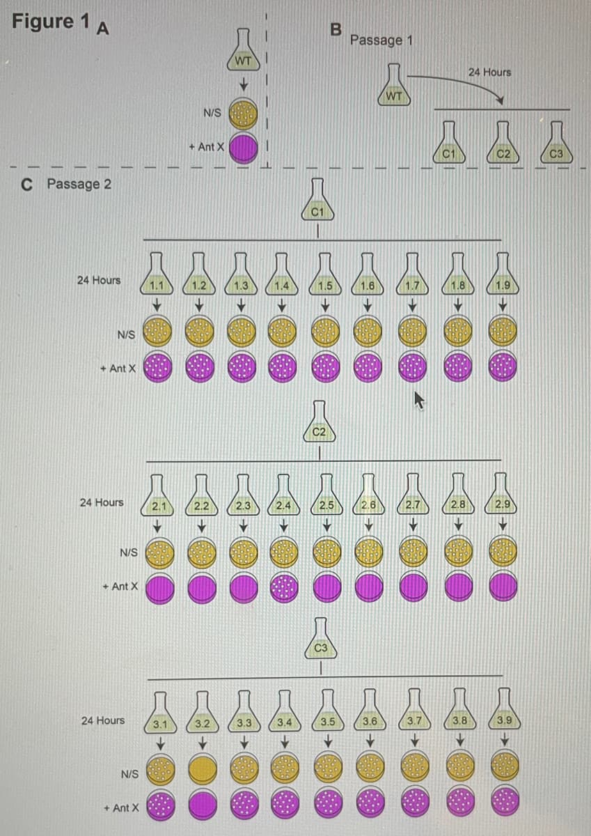
1) Analyse the following experimental set-up (illustrated in Figure 1) a) A wild-type (WT) bacterial strain is sensitive to the antibiotic X (Ant X) (Figure 1A) b) You have grown the WT strains in 3 independent liquid cultures (C1, C2 and C3) for 24 hours (Figure 1B-Passage 1) c) Following this initial growth, C1, C2 and C3 cultures are subcultured in 9 independent cultures and grown for 24 hours (Figure 1C Passage 2). Subsequently, cultures are plated on Non-Selective (N/S) solid media and media with Ant X (+ Ant X). The number of colonies grown in each plate are shown (Figure 1C) Use the above information to discuss: a. the type of mutations (if any) that have been arisen during the experiment. b. When did the mutations arise


Microscopic examination
The analysis of minute organisms, cellular organization of any biological structure, and composition of body fluids with the help of a microscope is known as microscopic examination. The magnification of specimens or samples under study helps in attaining a clearer picture of it.
Gram Staining
Named after Hans Christian Gram, a Danish bacteriologist, Gram stain is one of the most powerful staining techniques within microbiology. This technique was introduced in 1882 to identify pneumonia-causing organisms. The Gram staining technique uses crystal violet or methylene blue as primary staining colors to distinguish gram-positive from gram-negative organisms. Under a microscope, the gram-positive organisms appear purple-brown, retaining the primary color. Gram-negative organisms appear pink or red as they do not acquire the color of the primary stain.

Step by step
Solved in 3 steps


