Can I get help on how to do a concept map to the introduction of the Welch-Reardon, et al article provided below in the image. It can either be a drawing or a ppt slide. A requirement would be to utilize connecting words and identify the background of the article. Also, to research the problem and questions.
Can I get help on how to do a concept map to the introduction of the Welch-Reardon, et al article provided below in the image. It can either be a drawing or a ppt slide. A requirement would be to utilize connecting words and identify the background of the article. Also, to research the problem and questions.

Biochemistry
6th Edition
ISBN:9781305577206
Author:Reginald H. Garrett, Charles M. Grisham
Publisher:Reginald H. Garrett, Charles M. Grisham
Chapter32: The Reception And Transmission Of Extracellular Information
Section: Chapter Questions
Problem 21P

Related questions
Question
Can I get help on how to do a concept map to the introduction of the Welch-Reardon, et al article provided below in the image. It can either be a drawing or a ppt slide. A requirement would be to utilize connecting words and identify the background of the article. Also, to research the problem and questions.
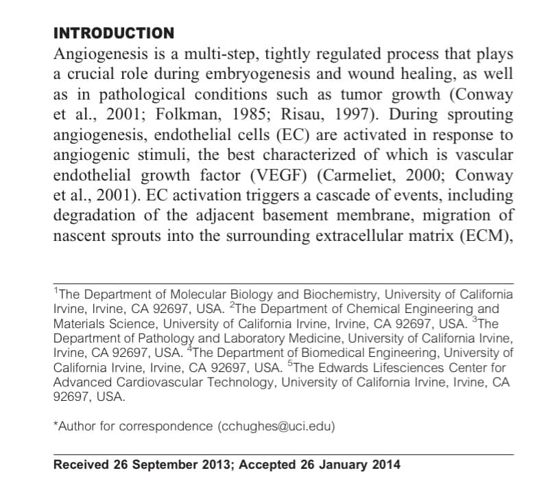
Transcribed Image Text:INTRODUCTION
Angiogenesis is a multi-step, tightly regulated process that plays
a crucial role during embryogenesis and wound healing, as well
as in pathological conditions such as tumor growth (Conway
et al., 2001; Folkman, 1985; Risau, 1997). During sprouting
angiogenesis, endothelial cells (EC) are activated in response to
angiogenic stimuli, the best characterized of which is vascular
endothelial growth factor (VEGF) (Carmeliet, 2000; Conway
et al., 2001). EC activation triggers a cascade of events, including
degradation of the adjacent basement membrane, migration of
nascent sprouts into the surrounding extracellular matrix (ECM),
"The Department of Molecular Biology and Biochemistry, University of California
Irvine, Irvine, CA 92697, USA. 2The Department of Chemical Engineering and
Materials Science, University of California Irvine, Irvine, CA 92697, USA. The
Department of Pathology and Laboratory Medicine, University of California Irvine,
Irvine, CA 92697, USA. “The Department of Biomedical Engineering, University of
California Irvine, Irvine, CA 92697, USA. SThe Edwards Lifesciences Center for
Advanced Cardiovascular Technology, University of California Irvine, Irvine, CA
92697, USA.
*Author for correspondence (cchughes@uci.edu)
Received 26 September 2013; Accepted 26 January 2014
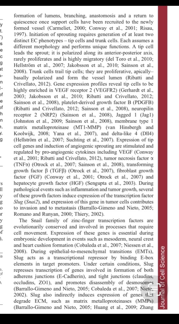
Transcribed Image Text:formation of lumens, branching, anastomosis and a return to
quiescence once support cells have been recruited to the newly
y
formed vessel (Carmeliet, 2000; Conway et al., 2001; Risau,
t.
1997). Initiation of sprouting requires generation of at least two
distinct EC phenotypes – tip cells and trunk cells. Each assumes a
different morphology and performs unique functions. A tip cell
leads the sprout; it is polarized along its anterior-posterior axis,
e
rarely proliferates and is highly migratory (del Toro et al., 2010;
g
Hellström et al., 2007; Jakobsson et al., 2010; Sainson et al.,
at
2008). Trunk cells trail tip cells; they are proliferative, apically-
basally polarized and form the vessel lumen (Ribatti and
Crivellato, 2012). Gene expression profiles reveal tip cells to be
highly enriched in VEGF receptor 2 (VEGFR2) (Gerhardt et al.,
of
2003; Jakobsson et al., 2010; Ribatti and Crivellato, 2012;
Sainson et al., 2008), platelet-derived growth factor B (PDGFB)
(Ribatti and Crivellato, 2012; Sainson et al., 2008), neuropilin
receptor 2 (NRP2) (Sainson et al., 2008), Jagged 1 (Jag1)
(Johnston et al., 2009; Sainson et al., 2008), membrane type 1
e,
e
of
h
matrix metalloproteinase (MT1-MMP) (van Hinsbergh and
Koolwijk, 2008; Yana et al., 2007), and delta-like 4 (Dl14)
(Hellström et al., 2007; Suchting et al., 2007). Expression of tip
cell genes and induction of angiogenic sprouting are stimulated and
regulated by pro-angiogenic cytokines including VEGF (Conway
et al., 2001; Ribatti and Crivellato, 2012), tumor necrosis factor a
(TNF¤) (Otrock et al., 2007; Sainson et al., 2008), transforming
growth factor B (TGFB) (Otrock et al., 2007), fibroblast growth
factor (FGF) (Conway et al., 2001; Otrock et al., 2007) and
hepatocyte growth factor (HGF) (Sengupta et al., 2003). During
pathological events such as inflammation and tumor growth, several
of these growth factors induce expression of the transcription factor
Slug (Snai2), and expression of this gene in tumor cells contributes
to invasion and to metastasis (Barrallo-Gimeno and Nieto, 2005;
Romano and Runyan, 2000; Thiery, 2002).
The Snail family of zinc-finger transcription factors are
evolutionarily conserved and involved in processes that require
of
ar
cell movement. Expression of these genes is essential during
embryonic development in events such as mesoderm, neural crest
and heart cushion formation (Cobaleda et al., 2007; Niessen et al.,
2008). During epithelial-to-mesenchymal transitions (EMTS),
Slug acts as a transcriptional repressor by binding E-box
elements in target promoters. Under certain conditions, Slug
represses transcription of genes involved in formation of both
adherens junctions (E-Cadherin), and tight junctions (claudins,
occludins, ZO1), and promotes disassembly of desmosomes
(Barrallo-Gimeno and Nieto, 2005; Cobaleda et al., 2007; Niete,
2002). Slug also indirectly induces expression of genes th.et
degrade ECM, such as matrix metalloproteinases (MMPS)
(Barrallo-Gimeno and Nieto, 2005; Huang et al., 2009; Zhang
ia
of
Journal of Cell Science
Expert Solution
This question has been solved!
Explore an expertly crafted, step-by-step solution for a thorough understanding of key concepts.

This is a popular solution!

Trending now
This is a popular solution!

Step by step
Solved in 2 steps with 4 images

Recommended textbooks for you
Biochemistry
Biochemistry
ISBN:
9781305577206
Author:
Reginald H. Garrett, Charles M. Grisham
Publisher:
Cengage Learning
Biochemistry
Biochemistry
ISBN:
9781305577206
Author:
Reginald H. Garrett, Charles M. Grisham
Publisher:
Cengage Learning